Download
1 / 29
570 likes | 1.96k Vues
Measurement Of Haematocrit (PCV). Dr Maliha Sumbul. Haematocrit:. Relative volume of blood occupied by erythrocytes. An average figure for humans is 45ml per cent, i.e. A packed red cell volume of 45ml in 100ml of blood. PCV / Packed cell Volume / Haematocrit.

E N D
Measurement Of Haematocrit (PCV) Dr Maliha Sumbul
Haematocrit: • Relative volume of blood occupied by erythrocytes. • An average figure for humans is 45ml per cent, i.e. A packed red cell volume of 45ml in 100ml of blood.
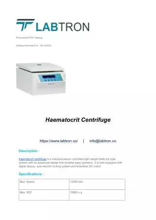
PCV / Packed cell Volume / Haematocrit High Speed Haematocrit centrifuge
Hematocrit (Ht or HCT) or Packed cell volume (PCV) or Erythrocyte volume fraction (EVF) • Proportion of blood volume that is occupied by red blood cells • It is normally about 46% for men and 38% for women • It is considered an integral part of a person's complete blood count results, along with hemoglobin concentration, white blood cell count, and platelet count • In mammals, hematocrit is independent of body size • The term "haematocrit" (British English: haematocrit) was coined in 1903. Its roots stem from the Greek words hema (Gr αίμα) - blood, and krites (Gr κριτής), judge - meaning to gauge or judge the blood.
Can be determined by centrifugingheparinized blood in a capillary tube (also known as a microhematocrit tube) at 10,000 RPM for five minutes • This separates the blood into layers • The volume of packed red blood cells, divided by the total volume of the blood sample gives the PCV • Because a tube is used this can be calculated by measuring the lengths of the layers.
With modern lab equipment, the hematocrit is calculated by an automated analyzer and not directly measured • It is determined by multiplying the red cell count by the mean cell volume • The hematocrit is slightly more accurate as the PCV includes small amounts of blood plasma trapped between the red cells • An estimated hematocrit as a percentage may be derived by tripling the hemoglobin concentration in g/dL and dropping the units
There have been cases where the blood for testing was inadvertently drawn proximal to an intravenous line that was infusing packed red cells or fluids. In these situations, the hemoglobin level in the blood sample will not be the true level for the patient because the sample would contain a large amount of the infused material rather than what is diluted into the circulating whole blood. That is, if packed red cells are being supplied, the sample will contain a large amount of those cells and the haematocrit will be artificially very high. • Conversely, if saline or other fluids are being supplied, the blood sample would be diluted and the hematocrit will be artificially low.
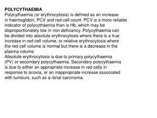
Elevated haematocrit • In cases of dengue fever a high hematocrit is a danger sign of an increased risk of dengue shock syndrome • Polycythemia vera (PV), a myeloproliferative disorder in which the bone marrow produces excessive numbers of red cells, is associated with elevated hematocrit • Chronic obstructive pulmonary disease (COPD) and other pulmonary conditions associated with hypoxia may elicit an increased production of red blood cells. This increase is mediated by the increased levels of erythropoietin by the kidneys in response to hypoxia • Professional athletes' hematocrit levels are measured as part of tests for blood doping or Erythropoietin (EPO) use; the level of hematocrit in a blood sample is compared with the long-term level for that athlete (to allow for individual variations in hematocrit level), and against an absolute permitted maximum (which is based on maximum expected levels within the population, and the hematocrit level which causes increased risk of blood clots resulting in strokes or heart attacks) • If a patient is dehydrated, the hematocrit may be elevated. Repeat testing after adequate hydration therapy will usually result in a more reliable result.
Lowered haematocrit • Lowered hematocrit can imply significant hemorrhage (for example, in an ectopic pregnancy) • The mean corpuscular volume (MCV) and the red cell distribution width (RDW) can be quite helpful in evaluating a lower-than-normal hematocrit, because it can help the clinician determine whether blood loss is chronic or acute. The MCV is the size of the red cells and the RDW is a relative measure of the variation in size of the red cell population. A low hematocrit with a low MCV with a normal RDW suggests a chronic iron-deficient erythropoiesis, but a high RDW suggests a blood loss that is more acute, such as a hemorrhage Groups of individuals who are at risk for developing anemia include: • infants who may not have adequate iron intake • children going through a rapid growth spurt, during which the iron available cannot keep up with the demands for a growing red cell mass • women in childbearing years who have an excessive need for iron because of blood loss during menstruation • pregnant women, in whom the growing fetus creates a high demand for iron. • patients with chronic kidney disease, as their kidneys no longer secrete sufficient levels of the hormone erythropoietin, which stimulates red blood cell production by the bone marrow.
REFERENCE VALUES • Varies with age and gender • At birth: 45 – 60% • At 1 yr: 27 – 44% • Adult: 36% – 48% for women 40 – 55% for men • Slight decrease after 50 yrs of age
MICROHAEMATOCRIT METHOD REAGENTS AND EQUIPMENT: • 1. Microhaematocrit Tube: 75 mm long, inner bore of 1.2 mm 2 types – heparinized tube (red band) -plain tube with anticoag. WB ( blue band ) The microhaematocrit tube holds approx. 0.05 ml of whole blood
Reagents and Equipment - continued • 2. Clay-like sealing compound • 3. Microhaematocrit centrifuge capable of producing RCF of 10,000 to 15,000 g. Also, should be able to reach max. speed within 30 sec. • 4. Microhaematocrit tube reader
SPECIMEN: • Whole blood using dipotassium EDTA as the anti-coagulant ( The liquid tripot. salts of EDTA can cause 2-3% decrease in haematocrit due to shrinkage of rbcs )
PRINCIPLE: • WHOLE BLOOD IS C/G FOR MAXIMUM RED CELL PACKING. THE SPACE OCCUPIED BY RBCS IS MEASURED AND EXPRESSED AS % OF WHOLE BLOOD VOLUME
PROCEDURE: • Allow the capillary or well-mixed WB to enter microht tubes until they are 2/3rds filled with blood (air bubbles denote poor technique but do not affect the results) • Seal one end of microht tube with clay material by placing the dry end in clay vertically (90 degree angle). The plug should be 4-6 mm long ( make certain that blood is not forced out during the process)
Place 2 microht. Tubes in the radial grooves of the C/G HEAD exactly opposite each other, with the sealed-end away from the centre of the centrifuge • C/G for 5 minutes • Remove the tubes as soon as the C/G has stopped spinning • Determine PCV with microht. Tube reading device
Duplicate results should agree within 1 unit (%) • If not, REPEAT THE PROCEDURE
PRECAUTIONS: • DO NOT INCLUDE BUFFY COAT WHEN READING RESULTS • AVOID PARALLAX ERROR ( PARALLAX: defined as an object being seen in a different position by changing the position of the head, or as seen by one eye versus the other eye)
UNIT OF EXPRESSION TWO WAYS: • As a %, e.g., 42% • As a decimal fraction, e.g., 0.42
Important to remember • Incomplete sealing – falsely low results due to escape of blood • Inadequate C/G or delay in reading results – falsely elevated results • Time and speed of C/G – V. important for max. red cell packing To detect: apply white tape to the inner side
Over-anticoagulated blood: HCT falsely low due to rbcs shrinkage • Trapped plasma: even with correct time period and proper speed - usually expressed as a % of the red blood cell - 1-3% higher b/c of trapped plasma when compared with automated cell counter - Increased amount of trapped plasma is found in: macrocytic anemias, spherocytosis, thalassemia, hypochromic anemias and sickle cell anemia
For accurate results, anticoagulated blood samples should be centrifuged within 6 hrs of collection when blood is stored at room temperature • Avoid heat sealing of the tubes - as flat sealing difficult to obtain and heat destroys rbcs
MACROHAEMATOCRIT METHOD • Old method • Rarely used now • More time-consuming • Requires larger amounts of blood • Contains higher % of trapped plasma METHOD: • Wintrobe tube, calibrated from 0 to 100 • Filled with anticoagulated blood and C/G at 2000 to 2300 g for 30 min • The ratio of the volume of rbcs to the total volumeof blood is then determined and reported as HCT reading