Download
1 / 43
430 likes | 502 Vues
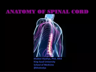
Gain insight on brain stem and spinal cord anatomy, functions, and pathways to understand the nervous system organization better. Discover motor pathways, reflexes, and sensory tracts. Dive into spinal nerve roots, motor neurons, and spinal reflexes. Explore dorsal columns and somatosensory system.

E N D
Basic Neuro AnatomyBrainstem and Spinal cord • G.Mathan Assistant Professor
Cerebrum Brain stem Midbrain Pons Medulla Cerebellum Brain and cerebellum
Brain Stem • three main parts • medulla, pons,midbrain. • Cranial nerves
Organization of the Nervous System • CNS • Brain • Spinal cord • PNS • Somatic • Autonomic • Sympathetic • Parasympathetic • Enteric
Functions of Spinal Cord • Final common pathway for the somatomotor system • Conveys somatosensory information from the body • Autonomic neurons • Reflexes • Central pattern generators for rhythmic movements and other fixed action patterns
Sensory vs motor roots “Bell and Magendie Law” Dorsal Root: Sensory (Afferents) Ventral Root: Motor (Efferents) Somatic motor & Visceral motor “The nerve which supply muscle groups also supply the skin over the joint movedby the muscles”
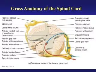
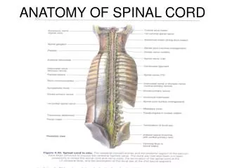
Internal Gross Anatomy • Anterior median fissure • Posterior median sulcus • Central canal • Anterior white commisure • Dorsal, ventral and lateral horn • Dorsal, ventral and lateral funiculus
Rexed’s Lamina • Some important nuclei • Substantia gelatinosa (II) • Nucleus proprius (IV) • Dorsal nucleus of Clarke • Intermediolateral cell column • Motor neuron pools
Alpha and gamma motor neurons • ventral horn cells, anterior horn cells • Very elaborate dendritic tree • Neurotransmitter=Ach • Alpha: extrafusal fibers • Gamma: intrafusal fibers • Motor pool = set of neurons that innervate a set or group of muscles • Motor unit: a motor neuron and its muscle cells (fibers) Motor Neurons
Muscle Spindles • Monitor stretch of muscle • Consist of intrafusal fiber, gamma motor fiber and sensory ending • Gamma motor neuron can alter sensitivity of muscle spindle www.utdallas.edu/.../ integ/mot1/display1_21.html
Motor Hierarchy(Swanson, 2003) • Central pattern controllers • Central pattern initiators • Central pattern generators • “Final common pathway”: motor pool • “As we move up the motor system hierarchy, away from the motoneuron pools themselves, explanations become more and more vague and the true situation in terms of neural networks becomes more and more complex.”
Motor Hierarchy • Lateral group (extremeties; fine motor control) • Corticospinal tract • Rubrospinal tract • Medial group (axial musculature; rhythmic and postural movements) • Vestibulospinal tract • Tectospinal tract • Reticulospinal tract • “Final common path”: motor pool
Corticospinal tracts • Also called pyramidal tract • Arises primarily from primary motor, premotor and supplementary motor cortex • Somatosensory cortex also contributes • 70-90% of fibers cross in the lower medulla (decussation of pyramids) • Crossed = lateral corticospinal tract • Uncrossed = anterior corticospinal tract • Synapses with: • Interneurons • Alpha and gamma motor neurons • Priopriospinal neurons
Spinal Reflexes • Programmed stereotypical reactions that occur in response to stimuli • Simplest reflex: monosynaptic stretch reflex • Examples of polysynaptic reflexes: withdrawal reflex • Reflexes are subject to higher level control
Submodalities are carried by different tracts • Dorsal columns • Fine, discriminitive touch, conscious proprioception, pressure and vibration sense • Facisculus gracilis vs fasciculus cuneatus • First synapse: relay nuclei in the medulla (nucleus gracilis and nucleus cuneatus) • Anterolateral system • Pain and temperature - First order neurons dorsal horn • Form Lissauer’s tract • Cross in anterior white commisure: ascend as spinothalamic tract (also spinoreticular and spinomesencephalic tracts) • SpinocerebellarTracts • Dorsal and ventral: ipsilateral • Unconscious proprioception
Organization of Somatosensory System • Dorsal Column : - • Gracilis & cuneate tracts - • Conscious proprioception , • Pressure and vibration, Fine discriminitive touch • Lateral Column :- • Lateral Spino thalamic Tract • Pain & Temperature • Posterior and anterior Spino cerebellar Tracts • Unconscious proprioception • Anterior Column:- • Anterior spinothalamic Tract • Crude touch, Itch and ticle sense
Dorsal Columns • Somatotopically organized • Medial Faciculus Gracilis • Lateral Faciculus Cuneatus • Conscious touch on contralateral side of the body • Concious proprioceptive sense • Two point tactile discrimation • Vibratiion and pressure
Dorsal Column/Medial Lemniscal system • Secondary neuron is in brainstem: • nucleus gracilis and • nucleus cuneatus • Output of these crosses midline and forms recognizable bundle: medial lemniscus • Medial lemniscus fibers synapse in the thalamus in the ventro posterior nuclei • Thalamic axons synapse in primary somatosensory cortex in several somatotopic maps with some segregation of submodalities http://cas.bellarmine.edu/tietjen/HumanBioogy/central_nervous_system.
Anterolateral system • Ist order of Neurons –Dorsal root ganglion • Run in Dorsolateral tract for few segements • Fibers thought to use substance P as a transmitter • Cross to form Lateral Spinothalamic, • Spinoreticular, & Spinomesencephalic tracts
Cerebral dominance Voluntary Movement and Cognitive Functions Lateralization = segregation of functions in the left and right Cerebral hemispheres of the brain Left hemisphere – language, math, logical operations, & serial processing of info sequences • Specializes in focused perception for the detailed, speed-optimized activities & the processing of fine visual and auditory details
Cerebral Dominance • Right hemisphere – pattern recognition, face recognition, spatial relations nonverbal thinking, emotional processing in general; the simultaneous processing of many kinds of info • Emphasis on understanding and generating stress & intonation patterns of speech that convey emotional content • Specializes in perceiving the relationship between images and the whole context in while they occur
Posterior and anterior Spino cerebellar Tracts Unconscious proprioception
Somatosensory Transduction • Sensory neurons in dorsal root ganglia • Touch • Prioprioception • Pain • Temperature • Pseudomonopolar • Use glutamate as a neurotransmitter • Also contain peptides, e.g., Substance P • Cell types have specialized endings for each modality • Encapsulated • (touch, proprioception) • Unencapsulated (free nerve endings) • Pain and temperature
Parasympathetic System • Generally associated with restorative functions, e.g., stimulation of peristaltic and secretory activities of the GI tract • Pre-ganglionic • Brainstem and sacral cord “Craniosacral division of ANS” • ACh as neurotransmitter • Post-ganglionic • Located either in the wall of the organs they innervate or in close proximity to target=more localized action • ACh as neurotransmitter (also peptides)
Sympathetic Nervous System • Mobilization of bodily resources • Flight or fight reactions • Pre-ganglionic neuron • spinal cord • ACh as transmitter • Post-ganglionic neuron • paravertebral chain=widespread action • Thoracic and lumbar regions of the cord=“Thoracolumbar division of ANS” • NE as neurotransmitter (except sweat glands = ACh) also peptides
Autonomic Ganglia Pre-ganglionic neuron (inside CNS) Post-ganglionic neuron (in PNS)
Acknowledgement • The Presentation is being used for educational and non commercial purpose • Thanks are due to all those original contributors and entities whose pictures • used for making this presentation.