Download
1 / 79
830 likes | 1.08k Vues
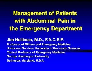
Management of Patients with Abdominal Pain in the Emergency Department. Jim Holliman, M.D., F.A.C.E.P. Professor of Military and Emergency Medicine Uniformed Services University of the Health Sciences Clinical Professor of Emergency Medicine George Washington University

E N D
Management of Patients with Abdominal Pain in the Emergency Department Jim Holliman, M.D., F.A.C.E.P. Professor of Military and Emergency Medicine Uniformed Services University of the Health Sciences Clinical Professor of Emergency Medicine George Washington University Bethesda, Maryland, U.S.A.
Abdominal Pain Lecture Outline • Recognition & resuscitation for life-threatening causes of abd. pain • Physical exam features • Choosing diagnostic tests • Initial treatment • Differential diagnosis • Key points about the most common specific causes
Abdominal Pain : Diagnostic & Treatment Priorities • First : recognize presence of shock or intraabdominal bleeding • Second : start resuscitative measures for shock or bleeding (if these are present) • Third : determine if the abdomen is the source of the shock or bleeding • Fourth : determine if emergency laparotomy is needed • Fifth : complete the secondary survey (head to toe exam) ; obtain needed lab or radiographic studies • Sixth : Conduct frequent reassessments of the patient
General Approach to the Patient Presenting with Abdominal Pain • Evaluate & treat the ABC's (Airway, Breathing, Circulation) first in same sequence as for any other emergency patient • Determine if an immediate life-threatening cause of abd. pain may be present & if there is any history of possible abd. trauma • Start resuscitation and emergently consult a surgeon if an emergent laparotomy is needed • Complete the secondary survey, treat pain, and decide what other diagnostic tests will be needed
Immediate Life-Threatening Causes of Abdominal Pain • These must be recognized from the primary survey : • Ruptured abdominal aortic aneurism (AAA) • Rupture of the spleen or liver • Ruptured ectopic pregnancy • Bowel infarction • Perforated viscus • Acute myocardial infarction (MI)
Ruptured Abdominal Aortic Aneurism (AAA) • More common in males > 65 years of age • May present initially as back or groin pain • Typically would have epigastric or periumbilical pain radiating to back • May present in shock from intraperitoneal rupture (retroperitioneal rupture may initially be contained) • Often can feel pulsating supraumbilical mass (if you can feel the aortic pulse width > 4 cm : suspect AAA) • Can sometimes make this Dx from lateral X-ray of abd. • Bedside ultrasound (U/S) is best Dx test for unstable patient • Abd. CT scan is best Dx test for stable patient (surgeon may also want angiography preop if patient is stable)
Emergency Management of Ruptured AAA • Oxygen & IV fluid resuscitation (normal saline or lactated Ringer's) if systolic BP < 100 mm Hg (but do not "overresuscitate" ; do not increase the BP to over 120 systolic because higher BP may cause increased bleeding) • Type and cross for at least 6 units of blood • Insert foley catheter • Obtain an electrocardiogram • Emergently consult a surgeon • Notify the operating room
Ruptured Spleen or Liver • Usually due to trauma, but can be spontaneous from malaria, mononucleosis, or hematologic diseases • Patient may present with shock ; may also have referred pain to shoulder (Kehr's sign) • Dx and Rx considerations & sequence same as for ruptured AAA (IV fluid, Type & cross, U/S or CT, call surgeon, etc.)
Ruptured Ectopic Pregnancy • Most common cause of pregnancy-related death in U.S.A. • May NOT have missed menstrual period • Typically have severe sudden onset lower abd. pain +/- shock • Should obtain stat serum or urine HCG test in any female of reproductive age with abd. pain • Pelvic U/S is Dx test of choice • Rx : Oxygen, IV fluid (NS or LR), Type & cross at least 2 units, emergently consult surgeon or obstetrician
Bowel Infarction • Due to clot embolus or thrombosis in mesenteric artery • Most patients have severe coronary artery disease (this can be a post-MI complication) • May have "pain out of proportion to findings" (may not demonstrate much tenderness) • Physical exam may show signs of peritonitis, hypoactive bowel sounds, blood in rectum or guiac positive stool
Bowel Infarction (cont.) • Usual lab findings : • High WBC • Severe lactic acidosis (anion gap > 18) • Plain X-ray film findings : • Free air, air in portal vein, air in bowel wall ("pneumatosis intestinalis") • May need emergent angiography for Dx • Rx : Oxygen, IV fluid resuscitation, IV broad spectrum antibiotics, consult surgeon
Angiogram (arrow shows superior mesenteric artery clot) of a 65 year old male with bowel ischemia
Perforated Viscus • Causes : • Blunt or penetrating trauma, tumors, inflammaory bowel disease, typhoid fever, amebiasis, other parasites • Typically see free air under diaphragm on plain films (Chest X-ray is most sensitive to see small amounts of air) • Rx : Oxygen, IV fluids, IV broad spectrum antibiotics (such as cefoxitin & metronidazole), emergently consult surgeon
Abdominal film showing the “Rigler double wall sign” of free intraperitoneal air (can see both inside and outside wall of bowel)
Acute Myocardial Infarction (MI) as a Cause of Abdominal Pain • Suspect in adult patient with upper abd. pain but no or minimal abd. tenderness • Inferior MI commonly presents as "indigestion" ; may also have emesis • MI may also secondarily occur from shock due to an intraabdominal cause (such as intraluminal bleed, etc.) • Dx by EKG +/- enzymes ; need Chest X-ray also • Rx : Oxygen, IV line, nitrates, aspirin, consider thrombolytics, etc., & admit to monitor bed unit
Now That Immediate Life-Threatening Causes of Abd. Pain Have Been Reviewed, Next the Lecture Will Review History and Exam for the Stable Patient • History items to ask the patient with abd. pain : • Time and rapidity of onset • Character of pain (burning, cramping, etc.) • Associated symptoms • Signs of bleeding (dark vomitus or stool) • Prior surgeries & illnesses • Last menstrual period • Medications (especially steroids, aspirin, warfarin) • Alcohol intake • Unusual ingestion or foreign travel
Physical Exam for the Patient with Abdominal Pain • Need complete set of vital signs • Look in nose and mouth for sites of bleeding (swallowed blood may mimic an intraluminal bleed) • Look at skin for stigmata of liver disease or signs of coagulapathy • Careful chest & lung exam (basilar pneumonias can present as abd. pain) • Palpate and observe the back • Genital and rectal exam (& stool guiac) should usually be routine
Exam of the Abdomen in the Patient with Abdominal Pain • Inspection : Look for : • Scars from prior surgeries • Distension • Localized swelling or mass • Eccymoses or erythema • Visible peristalsis • Auscultation with stethescope • Listen for bowel sounds & bruits • Palpation & percussion
Interpretation of Bowel Sounds (Associated, but not Definite, Diagnoses) • High pitched or "tinkling" : bowel obstruction • Continuous & hyperactive : acute gastroenteritis • Absent : ileus or peritonitis (need to listen for at least one minute) • Audible without stethescope : "borborygmi"
Percussion of the Abdomen • Should tap with 2 fingers on all 4 quadrants • If tympanitic : implies bowel obstruction • If dull, implies intraabdominal bleding or fluid (such as ascites) • If tender, correlate with tender areas noted on palpation
Palpation of the Abdomen • Should be done following inspection & auscultation • Assess for tenderness, guarding, mass, crepitus, referred tenderness • Differentiate lower rib tenderness from true upper abd. tenderness • Don't need to directly assess rebound ; just wiggle abdomen from the side & check for referred tenderness (direct rebound is cruel if peritonitis is present) • Don't forget leg maneuvers (psoas, obturator, & heel tap signs)
Lab Studies for Patients with Abdominal Pain • Use selectively ; not all are needed for all patients • For example, for young adults with simple acute viral gastroenteritis or food poisoning, usually no lab studies are needed (they may just need IV fluids & parenteral antiemetics) • Draw with the initial venipuncture if an IV line is to be established
List of Lab Studies to Consider for Patients with Abdominal Pain • Type and Cross (the most important if patient has shock) • Complete blood count (CBC) • Urine or serum pregnancy test (HCG) • Serum amylase, lipase • Urinalysis, urine culture and sensitivity • Liver function tests (bilirubin, SGOT, SGPT, alk. phos.) • Electrolytes, glucose, creatinine, blood urea nitrogen (BUN) • Serum alcohol, serum or urine drug screen • Serum medication levels (such as digoxin) • Clotting studies (platelet count, protime, PTT, fibrinogen) • Cardiac enzymes (if coronary ischemia suspected) • Blood culture (if sepsis or bacteremia suspected) • Nonemergent tumor markers (CEA, AFP)
Interpretation of Lab Studies for Abdominal Pain • WBC typically elevated (+/- "left-shifted") in any cause of peritonitis & in bowel infarction & in spleen & liver bleeding • However often NOT elevated appropriately in : • the elderly • immunocompromised patients • patients on chronic corticosteroid Rx
Interpretation of Lab Studies for Abdominal Pain (cont.) • Hematocrit may be normal in early stages of even severe hemorrhage • BUN to creatinine ratio of > 20 to 1 may indicate upper gastrointestinal (GI) bleed with digestion of blood in upper GI tract • Degree of elevation of amylase or lipase does not always correlate with severity of panceatitis or of pancreatic injury • Amylase may also be chronically elevated in patients with renal dysfunction
Plain Radiographs for Abdominal Pain • If needed, usually the 3 view "Acute Abdomen Series " is best (upright Chest X-ray, upright and flat plate of the abd.) • Chest X-ray best shows small amounts of free air • Upright abd. film best shows bowel air-fluid levels (indicating bowel obstruction or ileus if multiple) • Look also for abnormal calcifications • "KUB" film is oriented to include all the pelvis, whereas "abd. flat plate" is oriented to include the diaphragms (so these two are different for a tall patient)
Diagnostic Ultrasound for Abdominal Pain • Dx test of choice for : • Unstable patient in shock & suspected intraabdominal bleed • Gallstones (cholecystitis) • Ectopic pregnancy • Other complications of pregnancy (placenta previa, abruptio, etc.) • Renal or ureteral stones in the pregnant patient
Disadvantages of Diagnostic Ultrasound • Visualization may be limited by bowel gas or obesity • Good interpretation requires experience • Not good at showing retroperitoneal conditions • May not directly visualize solid organ lacerations
Use of Computed Tomography (CT) for Abdominal Pain • Noncontrast spiral scan is now method of choice for ureteral calculi (replaces intravenous pyelogram or IVP) • Using both IV and oral (or via nasogastric tube) contrast can then show appendicitis, diverticulitis, etc. • However even with greater use of CT for appendicitis, overall accuracy of this Dx in the E.D. has not improved
Other Diagnostic Studies to Consider for Abdominal Pain • If contrast CT not available : • Gastrografin Upper GI study for suspected : • Stomach or bowel perforation • Diaphragm rupture • Duodenal hematoma • Never do barium GI study if any chance of barium leak (causes severe peritonitis) • Intravenous pyelogram (IVP) for suspected : • Ureteral stone or injury • Renal mass
Other Diagnostic Studies to Consider for Abdominal Pain (cont.) • Retrograde urethrogram / cystogram for suspected urethral or bladder injury • Fistulogram for any suspected abdominal wall fistula • Technetium bleeding scan to localize intraluminal GI bleed • Angiography for preop planning of surgery for stable patient with AAA, or for suspected arterial bleed or mesenteric ischemia
Post-Exam "Procedures" to Consider for the Patient with Abdominal Pain • Insertion of foley catheter • Indicated for monitoring of any unstable patient or if urinary retention suspected • Insertion of nasogastric (NG) tube (see next slide) • Paracentesis (needle aspirate of abd. fluid) • Indicated for : • Suspected infected ascites (check cell count & culture) • Relieving tense ascites • Sometimes can make Dx of bowel perforation or intraabd. bleed
Usefulness Of NG Tube Suction for the Patient with Abdominal Pain • Allows decompression of stomach • Lessens risk of aspiration • Can remove some of residual toxins in stomach • May demonstrate upper GI bleeding • Required before peritoneal lavage • Contraindicated if nasal or midface fractures or severe coagulapathy (insert via mouth instead)
General Mechanisms Causing Abdominal Pain • Pain originating in the abdomen • Peritonitis • Distension of hollow viscera • Ischemia • Pain referred to the abdomen from another part of the body • Metabolic disorders • Neurogenic disorders
Causes of Referred Abdominal Pain from Chest Conditions • Acute coronary syndromes (and "angina equivalents") • Pneumonia (especially basilar) • Spontaneous pneumothorax • Pulmonary embolus (rare cause) • Pericarditis
Metabolic Causes of Abdominal Pain • Diabetic ketoacidosis • Hyperlipidemia (often with pancreatitis) • Acute prophyrias • Black Widow spider bites • Scorpion bites • Sickle cell crisis (sequestration in spleen or liver, or vaso-occlusive)
Neurogenic Causes of Abdominal Pain • Herpes zoster (Shingles) • Pain often present several days before characteristic dermatomal vesicles appear • Thoracic or lumbar spinal disc disease or compression • Syphilis ("tabetic crisis")
Trauma-Related Causes of Abdominal Pain • May present delayed, or from seemingly minor trauma in the elderly : • Ruptured spleen or liver • Bowel or stomach perforation • Pancreatic contusion or transection • Ruptured bladder • Mesenteric hematoma • Abdominal wall hematoma (U/S is good at diagnosing this)
Pregnancy-Related Causes of Abdominal Pain • Ectopic (usually tubal) pregnancy • False labor (Braxton-Hicks contractions) • Active labor • Abruptio placentae (note that placenta previa which can cause severe bleeding is usually painless) • Septic abortion
Genitourinary Tract Causes of Abdominal Pain • Cystitis • Pyelonephritis • Ureterolithiasis • Perinephric abscess (may see gas around kidney on KUB film) • Renal infarction (as from sickle cell disease) • Psoas abscess • Testicular torsion • Urinary retention (as from prostatic hypertrophy)