ANEMIAS
ANEMIAS. Dr. Aamer Aleem Consultant Hematologist Associate Prof. of Medicine KKUH & College of Medicine Riyadh. Anemia. Anemia is present when a patient has a reduced amount of hemoglobin per unit volume of blood when compared with the correct reference population for that patient.


ANEMIAS
E N D
Presentation Transcript
ANEMIAS Dr. AamerAleem Consultant Hematologist Associate Prof. of Medicine KKUH & College of Medicine Riyadh
Anemia • Anemia is present when a patient has a reduced amount of hemoglobin per unit volume of blood when compared with the correct reference population for that patient. • Males Hb-13-18 • Females Hb 12-17
ANEMIA • Single cell line(RBC) problem • Multiple cell line problem (RBC,WBC,Platelet) -Bone marrow suppression -immunologic disorders -peripheral destruction/sequestration
Anemia • Anemia is generally defined as a hematocrit <40% (hemoglobin <13.0 g/dL) in men or <37% (hemoglobin <12.0 g/dL) in women. (WHO definition) Red blood cell (RBC) indices, which include the mean corpuscular volume (MCV), the mean corpuscular hemoglobin (MCH), the mean corpuscular hemoglobin content (MCHC), and the red-cell distribution width (RDW) index, are further used to define types of anemia.
Anemia • Despite having a set of peculiar symptoms and signs, anemia is not a disease per se, but a syndrome, as it may arise from an extensive list of causes. • It is the chronic syndrome of highest prevalence in clinical medicine.
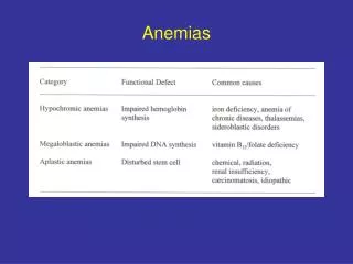
AnemiaEtiology • Based on Hb, red cell indices, retic count and red cell morphology • (1) Inadequate response A. Hypochromic microcyctic B. Normochromic Normocytic C. Macrocytic • (2)Adequate response R/O blood loss---Includes Hemolytic disorders
Anemia-symptoms • What are the symptoms of Anemia ? • General malaise, weakness, fatigue, breathlessness on exertion, palpitations, angina. • Desire to eat sand and clay. • Menorrhagia common in women.
Symptoms of Anemia • The central nervous system, the heart and the muscle mass are the most affected organs, since they are the ones that most need oxygen for their functions. • The symptoms increase with physical activity, as this consumes oxygen. • With hemoglobin between 9 and 11 g/dL there's irritability, headache and psychic asthenia; in the elder fatigability is observed, and angina may occur. • With hemoglobin between 6 and 9 g/dL there's tachycardia, dyspnea and fatigue upon the slightest effort. • With hemoglobin below 6 g/dL symptoms are present even in sedentary activities, and when below 3.5 g/dL heart failure is impending and any activity is unfeasible.
COMPLETE BLOOD COUNT • HbConc (g/dl) • Hematocrit (PCV) • MCV (fl) • MCH (pg) • RDW(measure of red cell size variability) • RBC Count (…x109/L ) • WbC Count (…x109/L ) • Platelet Count (…x109/L) • (Reticulocyte Count) ( % )
Red Cell Indices MCV defines the size of the red blood cells and is expressed as femtoliters (10−15; fl) or as cubic microns (μm3). The normal values for MCV are 87 ± 7 fl. MCH quantifies the amount of hemoglobin per red blood cell. The normal values for MCH are 29 ± 2 picograms (pg) per cell. MCHC indicates the amount of hemoglobin per unit volume. In contrast to MCH, MCHC correlates the hemoglobin content with the volume of the cell. It is expressed as g/dl of red blood cells or as a percentage value. The normal values for MCHC are 34 ± 2 g/dl.
Red Cell Indices How to calculate red cell indices
Glossary of Useful Blood Count Descriptors Anisocytosis: red cells of unequal size. Reflected in increased RDW (Red cell Distribution Width.) Dimorphic Blood Film: two populations of red cells - one microcytic and the other normocytic. Seen in treated or transfused iron deficiency, and sideroblastic anemia Howell-Jolly bodies: round nuclear remnants within the red cells. Indicate splenectomy or hyposplenismMacrocytosis: large red cells Erythroblast: any nucleated red cell precursor Hypersegmented neutrophils: a neutrophil with six or more lobes. Usually (but not inevitably) means vitamin B12 or folate deficiency
Glossary of Useful Blood Count Descriptors Hypochromia: pale red cells. Always accompanied by microcytosis Leukoerythroblastic: the presence of erythroblasts and myelocytes (which are precursors of mature cells) in the blood. Often indicates marrow infiltration eg by secondary cancer or fibrosis Microangiopathy: indicates mechanical damage to red cells with red cell fragments on the blood filmMicrocytosis: small red cells Poikilocytosis: a traditional term for red cells of unequal shape Polychromasia: grey coloured red cells on film, indicating presence of increased reticulocytes Reticulocyte: an erythrocyte newly released from the bone marrow
Glossary of Useful Blood Count Descriptors • Rouleaux: red cells in stacks, as coins. Indicates high ESR, eg infection, myeloma, cancer, collagen disease, TB etc. • Schistocyte: a red cell which has undergone mechanical damage - synonymous with red cell fragment • Spherocyte: a spherical red cell due to disproportionate membrane loss. Either inherited, or acquired from (usually) immune causes • Sickle cell: a crescent-shaped red cell characteristic of Sickle Cell Anemia • Target cell: red cell with central area of Hb giving the appearance of a target. Seen in many conditions, including IDA, hemoglobinopathy and liver disease
Rouleaux (singular is rouleau) are stacks of red blood cells. They occur when the plasma protein concentration is high, and because of them the ESR (erythrocyte sedimentation rate) is also increased. Conditions which do this include infections, inflammatory and connective tissue disorders, and cancers. In this case, the diagnosis is probably multiple myeloma, as a plasma cell can be seen on the film. Rouleaux
Erythroblasts are the precursor cells of erythrocytes. They contain nuclei, and in adults are only found in the bone marrow under normal circumstances. Their presence in the blood may indicate either marrow 'stress', as in hypoxia, severe sepsis, or hemolysis, for example, or some fundamental bone marrow pathology such as replacement by secondary cancer. Erythroblasts & other BM cells
Anisocytosis means that the red cells are of unequal size. It is a feature of many anemias, and other blood conditions, and does not have much diagnostic value. The 'red cell distribution width' (RDW) is a quantitative measure of the degree of anisocytosis. The RDW is useful in the differential diagnosis of microcytic anemia. Most cases of iron deficiency have a raised RDW, and most cases of thalassemia trait have a normal RDW. Anisocytosis
There are two populations of red cells present. One is normocytic, and the other is microcytic. This occurs either because an iron-deficient patient has been transfused or treated with iron, or in the Sideroblastic Anemias. Dimorphic Red Blood Cells
Anemia-diagnosis • How can we diagnose Anemia ? Blood tests to detect the various components of blood is carried out for proper diagnosis. Red blood cell count may be normal or decreased. Peripheral blood smear shows pale small cells. White blood cell count normal or decreased
Classification of anemia • Anemia can be either acute or chronic. • In acute anemia (sudden loss of blood), the lack of blood volume in the circulatory system is more important than the deficiency of hemoglobin. A loss up to 10% of blood volume, as that taking place upon blood donation, is well tolerated. Losses between 10 and 20% cause postural hypotension, dizziness and faint. In losses above 20%, there's tachycardia, cold extremities, extreme paleness and hypotension, followed by shock; should the loss surmount 30%, without immediate replacement of intravenous fluids, the shock rapidly becomes irreversible and fatal. • In chronic anemia, there's no decrease in blood volume, which is compensated by an increase in plasma volume.
Classification of anemia Based on MCV • Microcytic anemia • Normocytic anemia • Macrocytic anemai
Classification of anemia Based on underlying process • Blood loss or def. of nutrients • Hemolysis • Failure of production
A diagnostic scheme for anemias Determine the MCV • The most useful initial approach to anemia is based on red cell size. The Mean Corpuscular Volume (MCV) represents a direct measurement of red cell size. When this is reduced, the anemia is referred to as microcytic. It is macrocytic when the MCV is increased, and normocytic when the MCV is normal.
Anemia-classification • One way to classify anemia is by RBC size (i.e., MCV), as microcytic, macrocytic, or normocytic. For the microcytic anemias, the etiologic possibilities are iron deficiency, thalassemia, sideroblastic anemia, and the anemias of chronic disease. Severe microcytic anemia (MCV <70 fL) is caused mainly by iron deficiency or thalassemia. Macrocytic anemia may be the result of megaloblastic (folate or vitamin B12 deficiency) or nonmegaloblastic causes. Folate deficiency can in turn be due to either reduced intake or diminished absorption. Severe macrocytic anemia (MCV >125 fL) is almost always megaloblastic. In some rare cases, macrocytic anemia is related to the myelodysplastic syndromes prior to or after chemotherapy. • The causes of normocytic anemias include aplastic anemia, bone-marrow replacement, pure red-cell aplasia, anemias of chronic disease, hemolytic anemia, and recent blood loss. A number of anemias have a genetic etiology. Examples of such inherited disorders include hereditary spherocytosis andsickle-cell (SC) anemia
Hypochromic Microcytic AnemiaD/D Microcytic anemias the etiologic possibilities are • Iron deficiency • Thalassemia • Sideroblastic anemia • Anemias of chronic disease. Severe microcytic anemia (MCV <70 fL) is caused mainly by iron deficiency or thalassemia.
Normochromic Normocytic AnemiaD/D • Chronic inflammatory disease—(1)infection (2)collagen vascular disease (3)inflammatory bowel disease • Recent blood loss • Malignancy/Marrow infiltration • Chronic renal failure • Transient erythroblastopenia of chidhood • Marrow aplasia/hypoplasia • HIV infection • Hemophagocytic syndrome
Macrocytic Anemia D/D • Megaloblastic anemias • Vit.B12 def. - (1) pernicious anemia (2) malabsorption • Folate def. - (1) malnutrition (2) malabsorption (3) chronic hemolysis (4)drugs - phenytoin, sulfa • Hemolysis • Myelodysplastic syndrome • Marrow failure - Aplastic anemia • Chronic liver disease • Hypothyroidism
Macrocytic anemia D/D • Macrocytic anemiamay be the result of megaloblastic (folate or vitamin B12 deficiency) or nonmegaloblastic causes. Folate deficiency can in turn be due to either reduced intake or diminished absorption. Severe macrocytic anemia (MCV >125 fL) is almost always megaloblastic.
The Normocytic Anemias These may be classified as follows: • underproduction of erythrocytes due to (1) the anemia of chronic disease (2) marrow failure (3) renal failure (decreased erythropoietin) • loss or destruction of erythrocytes due to (1) hemolysis (2) acute blood loss • The reticulocyte count is useful in drawing this distinction, being elevated in (b) and reduced in (a).
The Normocytic Anemias • The causes of normocytic anemias include aplastic anemia, bone-marrow replacement, pure red-cell aplasia, anemias of chronic disease, hemolytic anemia, and recent blood loss. A number of anemias have a genetic etiology. Examples of such inherited disorders include hereditary spherocytosis, sickle-cell (SC) anemia, and thalassemia
Hemolytic Disorders • Hemoglobinopathy– Hb SS,SC,S-Bthal • Enzymopathy--G6PD def, PK def • Membranopathy—Hereditary spherocytosis, elliptocytosis • Extrinsic factors—DIC, TTP, HUS, • Immune hemolytic anemia---Autoimmune, Drug induced
Diagnostic Approach-History • Age : Iron def rare without blood loss before 6 months in term infants. • Family Hist &Genetics: (1)X-linked: G6PD deficiency (2)Aut dominant: Spherocytosis (3)Aut recessive:Sickle cell,Fanconi anemia (4)Family member with early age of cholecystectomy/splenectomy (5)Ethnicity (Thalassemia: Mediterranean; G6PD def: (Greeks, Blacks, Middle eastern) (6)Race:B-thal:Mediterranean,African,Asian; A-thal: Blacks, Asians
Diagnostic Approach-History • Nutrition: (1)Cows milk diet:iron def. (2)Strict vegetarian:Vit B12 def. (3)Goats milk: Folate def. (4)Pica: Plumbism,Iron def. (5)Cholestasis, malabsorption:Vit E def • Drugs: (1)G6PD: oxidants (sulfa, primaquine) (2)Immune mediated hemolysis(penicillin) (3)Bone marrow suppression(chemotherapy) (4)Phenytoin increases folate requirement
Diagnostic Approach-History Diarrhoea-Malabsorption of VitB12/E/Fe. Inflammatory bowel disease and anemia of chronic disease with or without blood loss. Milk protein intolerance induced blood loss Intestinal resection: Vit B12 def Infection: Giardia:iron malabsorption Intestinal bacterial overgrowth:Vit B12 def Fish tapeworm:Vit B12 def -EBV,CMV,Parvovirus:BMsuppression Mycoplasma,Malaria:hemolysis Hepatitis:aplastic anemia Endocarditis, HIV
Physical exam reveals presence and potential causes of anemia • Fever-acute infection,intravascular disease,collagen vascular disease • Jaundice suggests hemolysis • Petechia & Purpura—bleeding tendency • Hypertension & edema-renal disease • Hepatosplenomegaly and lymphadenopathy—infiltrative disease • Growth failure or poor wt. gain—Anemia of chronic disease or organ failure • Examine stool for blood; urine for hemoglobinuria (Admit & tranfuse if signs of CHF)
Physical Findings in Anemia • Skin: Hyperpigmentation, café-au-lait spots- Fanconi anemia Petechia & purpura-BM infiltration, autoimmune hemolysis & thrombocytopenia Erythematous rash-Parvovirus, EB virus Butterfly rash-SLE ; Vitiligo-Vit B12 def. • Head:Frontal bossing-Thalassemia major, Microcephaly-Fanconi anemia • Mouth: Glossitis-B12 def, iron deficiency Angular stomatitis-Iron deficiency Pigmentation-Peutz Jeghers syndrome Telangiectasia-Osler Weber Rendu syndrome
Physical Findings in Anemia • Extremities:Absent thumb-Fanconi anemia -Spoon nails-Iron deficiency -Dystrophic nails-Dyskeratosis congenita • CNS-Irritable, apathy-Iron def. -Peripheral neuropathy-lead poisoning -Ataxia, post.column signs-Vit B12 def - -Stroke-Sickle cell anemia • Short stature-Fanconi anemia, Malnutrition
Laboratory Evaluation • Hematology: Complete Blood Count, Retic count, Peripheral smear, ESR, G6PD, Sickling(+/- inf), Hb electrophoresis, Coombs Test, Osmotic fragility test, BM aspiration • Biochemistry: LFT, UE, RFT, S.Ferrtin, S.Haptoglobin, Iron, VitB12, Folate, Ceruloplasmin • Serology:Heterophil antibody, ANA,Viral • Urinalysis, microscopy, culture/sensitivity • Stool exam. for ova, parasites, occult blood • Endoscopy: upper and lower bowel • Imaging: US Abdomen, Skeletal radiographs, Tc pertechnetate scan for Meckels diverticulum • Tissue biopsy: skin, lymph node, liver
Laboratory Evaluation • Hematology: Complete Blood Count, Retic count, Peripheral smear, ESR, G6PD, Sickling(+/- inf), Hb electrophoresis, Coombs Test, Osmotic fragility test, BM aspiration • Biochemistry: LFT, UE, RFT, S.Ferrtin, S.Haptoglobin, Iron, VitB12, Folate, Ceruloplasmin • Serology:Heterophil antibody, ANA,Viral • Urinalysis, microscopy, culture/sensitivity • Stool exam. for ova, parasites, occult blood • Endoscopy: upper and lower bowel • Imaging: US Abdomen, Skeletal radiographs, Tc pertechnetate scan for Meckels diverticulum • Tissue biopsy: skin, lymph node, liver
Microcytic Anemia • TEST Iron def ThalMin An of ch dis • S.Iron - low normal normal • S.Ferritin - low N/H N/H • Marrow iron - low N/H N/H • Hb A2 or F - N H=Bthal N • N=Athal • RDW - high normal N/H • MCV ÷ RBC - >13< 13 • Sickle/B-thal – Hb S > Hb A • Absence of microcytosis in both parents excludes B-thal or Sickle/B-thal but not A-thal
Anemia-treatment How can Anemia be treated ? As there are many factors causing anemia, the main treatment is for the cause. • Specific treatment of disorder causing anemia. • Replacement of missing factors (iron, vitamin B12, folate) • In many chronic anemias regular blood transfusions are needed (thalassemia, myelodysplastic syndrome) • Blood transfusion is usually needed in acute blood loss or if the patient has severe symptoms or in heart failure • (Problems of blood transfusion)
Iron deficiency anemia (IDA) • It is a condition when supply of iron in the body to bone marrow falls short of that required for the production of red blood cells. It is the commonest cause of anemia throughout the world.
Iron Deficiency Anemia • The incidence of anemia in the general population is about 1.5%. • Iron deficiency related to inadequate replacement of lost iron is the most frequent cause of asymptomatic anemia and has a variety of causes. • Iron deficiency is common among women of childbearing age; 10% to 20% of menstruating women have abnormally low concentrations of hemoglobin (usually <12 g per 100 mL).
Iron Deficiency Anemia • What are the causes of IDA ? • Increased physiological demand for more red blood cells eg: increased physical activity. • In children, during spurts of growth. • In women during menstruation, pregnancy, parturition and lactation. • Inadequate dietary intake due to poor economic reasons or deficient foods. • Decreased absorption due to disorders in the digestive system. • GI blood loss Peptic ulcer, piles, hiatus hernia, carcinoma of stomach, carcinoma colon, chronic ingestion of a certain type of pain relievers, hookworm infestation.
Iron Deficiency Anemia Between 20% and 60% of pregnant women have hemoglobin levels <11 g per 100 mL. Anemia was found in 6% of white women and 17% of black women during the first trimester and in 25% of white women and 46% of black women during the third trimester.
Iron Deficiency Anemia The high rates of anemia in pregnant women are not attributable to iron deficiency alone, however. In a large study of low-income, urban, mostly minority pregnant women, only 12.5% were iron deficient. Women progressing through pregnancy develop a lowered hematocrit as a result of physiologic hydremia related to a disproportionate increase in plasma volume.
Iron Deficiency Anemia • The daily oral iron requirement for men and postmenopausal women is 0.5 and 1 mg • Menstruating and pregnant women have higher requirements: 2 mg and 2.5 mg, respectively. • The replacement of lost iron is complicated by the fact that only 5% to 10% of the 10 to 20 mg of the iron in the average adult diet is absorbed. • Anemia is less likely to occur in women taking birth control pills and more likely to occur in women with intrauterine devices.
Iron Deficiency Anemia Because men and postmenopausal women rarely develop iron deficiency that is not related to gastrointestinal blood loss (often occult), an evaluation of gastrointestinal tract must be performed when an iron deficiency is detected in these individuals