
Staining for acLDL and CRP in HUVECs: Effects of Preincubation with nCRP and mCRP
This study presents Supplemental Data Figure 2b, which examines the staining patterns of acLDL (a-c) and CRP (d-f) in Human Umbilical Vein Endothelial Cells (HUVECs) following preincubation with 0.1% fetal calf serum (FCS), 10 mg/L nCRP, and 10 mg/L mCRP. White arrows indicate the presence of LDL-containing vesicles, showing a reduction in mCRP-treated cells. Blue staining represents nuclear DNA (DAPI), while green shows nCRP (e) and mCRP (d, f). Cells treated with nCRP exhibited no staining, whereas mCRP treatment highlighted predominant membrane staining.

Staining for acLDL and CRP in HUVECs: Effects of Preincubation with nCRP and mCRP
E N D
Presentation Transcript
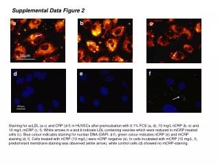
a c d 200µm 200µm b e f 30µm Supplemental Data Figure 2 b Staining for acLDL (a-c) and CRP (d-f) in HUVECs after preincubation with 0.1% FCS (a, d), 10 mg/L nCRP (b, e) and 10 mg/L mCRP (c, f). White arrows in a and b indicate LDL containing vesicles which were reduced in mCRP-treated cells (c). Blue colour indicates staining for nuclear DNA (DAPI, d-f), green colour indicates nCRP (e) and mCRP staining (d, f). Cells treated with nCRP (10 mg/L) were nCRP negative (e). In cells incubated with mCRP (10 mg/L, f), predominant membrane staining was observed (white arrow), while control cells (d) showed no mCRP-staining.