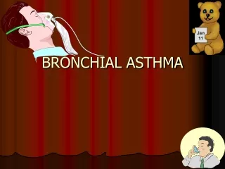
Bronchial asthma
Bronchial asthma. L de Man Dept of Physiotherapy UFS 2012. Definition. Asthma is a disease characterised by a wide variety of resistance to airflow

Bronchial asthma
E N D
Presentation Transcript
Bronchial asthma L de Man Dept of Physiotherapy UFS 2012
Definition • Asthma is a disease characterised by a wide variety of resistance to airflow in the intrapulmonary airways. This can occur in the absence of any other disease that can cause it and is reversible spontaneously or with medication
Aetiology and epidemiology • Has a genetic component • Tendency to allergies are inherited • Allergens : • faeces of house mite, • fur of cats, dogs, other animals, • grass pollens • certain foods • Also exercise, air temperature changes , paints, glues, NSAID, ß-blockers, stress, emotional disturbances. • Respiratory tract infection can also trigger attack
Aetiology and epidemiology Extrinsic Intrinsic • Earlier in life – childhood • Positive family history • Hypersensitivity to allergens • Positive skin-prick test • Occurs intermittent • Improves with age • Seasonal rhinitis and eczema • Occurs later in life • No family history • Allergic features absent • No positive skin-prick test • Occurs persistently • Worsens with age • Commoner in women
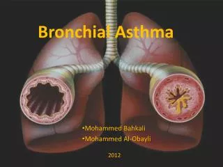
Pathophysiology • Obstruction occurs in the airways due to • Hypertrophy and hyperplasia of the bronchial smooth muscles • Thickening of the epithelial basement membrane of the airways • Oedema of the bronchial wall • Eosinophilic infiltration of the bronchial wall • Hypertrophy of the bronchial mucous glands • increase in number of goblet cells
Pathophysiology • Leads to narrowing of larger bronchi and plugging of bronchi and bronchioles with viscid mucus
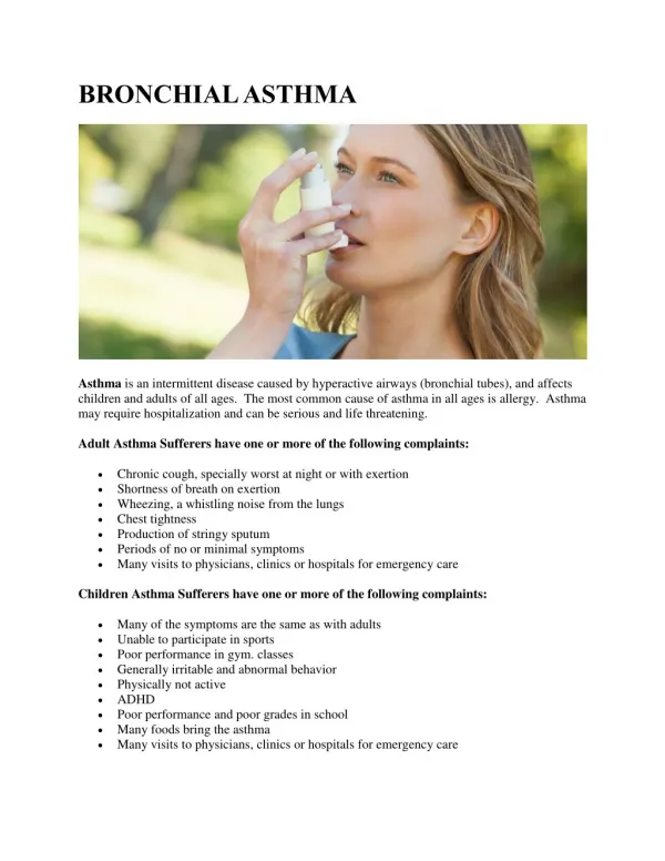
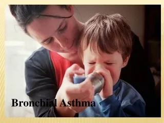
Clinical features • Wheeze • Breathlessness • Chest tightness • Cough that may be paroxysmal • Respiratory rate increased • Expiration prolonged • Use of accessory muscles of breathing • Decreased exercise tolerance
Medical management Preventers Relievers • Anti-inflammatory drugs (inhaled steroids)to suppress underlying inflammation • Avoidance of allergens! • Drugs that relieve bronchospasm • Inhaled ß2-agonists and anti-cholinergic drugs
Chest x-ray • Will show signs of hyperinflation with an acute attack
Status Asthmaticus • Acute, severe asthma severe wheezing and breathlessness lasting more than 24 hours • Not responding to normal medication. • Potentially life-threatening • Respiratory rate > 25/minute • Tachycardia > 110 bpm • Silent chest • Cyanosis • Disturbance in consciousness
Physiotherapy problems • Decreased airflow due to bronchospasm • Dyspnea due to decreased airflow • Tense shoulder girdle due to use of accessory muscles of breathing • Decreased exercise tolerance
Physiotherapy treatment • Relieve bronchospasm with inhalation therapy • Dyspnea management – relaxation positions, shoulder girdle relaxation, controlled breathing (relaxed diaphragmatic breathing, inspration and expiration relaxed, expiration prolonged), FET. • Increase exercise tolerance • Correct use of inhaler • Education • Use of peak flow meter for self management
Peak flow meter • Measures the fastest rate of airflow with forced expiration in l/min • Patient can use his predicted versus actual reading to indicate the need for treatment • Measure peak flow every day • If reading 60% of personal best, go to doctor • If reading ‹ 60% of personal best, go to emergency room
Peak flow meter • Allow one trial attempt to familiarise with device • Deep breathe in, device between lips, keep device level, seal lips tightly around device, blow out as hard as possible. • Take 3 readings immediately after each other • Record best of 3 readings. • See chart to determine predicted peak flow rate for the individual
References Downie,P.A. 1992. Cash’s Textbook of Chest, Heart and Vascular disorders for Physiotherapists. 4th ed. Mosby , 458-463; 507-511. Smith,M. & Ball, V. 1998. Cardiovascular/ Respiratory Physiotherapy. Mosby,171-174.