Download
1 / 36
380 likes | 921 Vues
Hemolytic Anemias due to Other Intracorpuscular Defects. What is intrinsically wrong with this RBC?. Other Intracorpuscular Defects.

E N D
Hemolytic Anemias due to Other Intracorpuscular Defects What is intrinsically wrong with this RBC?
Other Intracorpuscular Defects • Hereditary membrane defects – abnormalities in the constituent membrane proteins or lipids may alter the function and/or flexibility, shape, or stability of the membrane leading to hemolysis, most of which is extravascular via the spleen.
Intracorpuscular Defects • Hereditary spherocytosis (HS) • This is a group of heterogenous disorders which usually have an autosomal dominant inheritance (deletion in short arm of chromosome 8). • The specific defect is a disorder of vertical protein interactions, most often characterized by a deficiency of spectrin. • The deficiency may be a primary deficiency in spectrin or a secondary deficiency due to defective attachment of the cytoskeleton to the lipid bilayer. • Deficiencies in band 3, ankyrin and protein 4.2 have also been found
Intracorpuscular Defects • The net result of the skeletal defect is increased membrane instability and progressive membrane loss. • This leads to a cell with a decreased surface to volume ratio and spherocytes with decreased flexibility and increased viscosity which all leads to extravascular hemolysis. • The cells may also be more permeable to sodium and this eventually leads to decreased potassium and water and increased cell viscosity which also contributes to splenic culling. • Clinical manifestations: • Jaundice, mild to severe anemia, and an enlarged spleen.
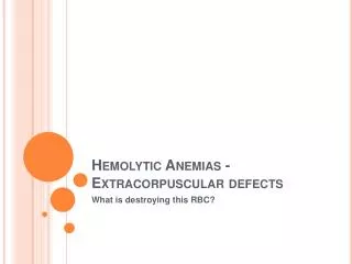
Intracorpuscular Defects • May have gallstones from the increased bilirubin. • The MCHC is increased (greater than 36%). • The peripheral smear shows spherocytes and polychromasia • There is increased osmotic fragility • The autohemolysis test is positive. In this test RBCs are incubated in their own plasma for 48 hours at 370 C and the amount of hemolysis is determined. • Normal is .2-2%. • With hereditary spherocytosis, the hemolysis is 5-25% at 24 hours, and 75% at 48 hours. • The hemolysis will decrease when glucose is added for making ATP to pump out excess Na.
OSMOTIC FRAGILITY TEST Fresh blood Incubated blood
Intracorpuscular Defects • Therapy = splenectomy • Hereditary elliptocytosis (HE) • This disease usually has an autosomal dominant inheritance • Several subgroups which vary in the degree of hemolysis and clinical severity are found. • There appears to be a spectrin dimer-dimer association problem. • Decreased amounts of protein 4.1 have also been found • The defect in the membrane cytoskeleton that results causes the abnormal shape (elliptocytes or ovalocytes).
Intracorpuscular Defects • In the heterozygote form the weakened skeleton results in cells being permanently deformed when subjected to the sheer stress of the microcirculation (this is transient in normal RBCs). • The cells have a nearly normal life span. • In the homozygote state (and with some heterozygote variants) there is severe weakening of the cytoskeleton and membrane fragmentation occurs in addition to the formation of elliptocytes. • This is called a hemolytic variant. • Clinical manifestations: • For the heterozygote it is usually a mild, compensated anemia with elliptocytes and polychromasia. The osmotic fragility and autohemolysis tests are normal. • In the homozygous state (rare), there is marked poikilocytosis with elliptocytes and fragmented cells. There is increased osmotic fragility and autohemolysis.
Intracorpuscular Defects • Therapy - splenectomy in hemolytic variants • Hereditary pyropoikilocytosis (HPP) • This is a rare autosomal recessive disorder with unstable spectrin leading to an unstable membrane cytoskeleton that undergoes sheer stress in the microvasculature described above for homozygous elliptocytosis. • HPP cells actually have two spectrin defects, one from each parent. • There is a deficiency of alpha spectrin from one parent. • There is a mutant spectrin that prevents self-association of heterodimers from the other parent
Intracorpuscular Defects • This leads to a severe hemolytic anemia and marked poikilocytosis with budding cells, fragments, microspherocytes, and elliptocytes. • The cells are thermally unstable and fragment at 45-460 C, whereas normal cells fragment at 490 C. • There is increased osmotic fragility and autohemolysis.
Intracorpuscular Defects • Disorders of membrane cation permeability • Hereditary stomatocytosis (stoma= mouth) • In this disorder the RBC is abnormally permeable to sodium and potassium. • The net gain of sodium is more than the net loss of potassium. • Therefore water rushes in and the RBCs are swollen and less deformable and are culled in the spleen. • The anemia may be mild to moderate with polychromasia. • Stomatocytes (RBCS with slit-like or mouth-like areas of central pallor) are seen • There is increased osmotic fragility and autohemolysis.
Intracorpuscular Defects • Hereditary xerocytosis • In this disorder there is a net loss of potassium that exceeds the net gain of sodium resulting in a dehydrated cell as water rushes out. • This leads to increased cytoplasmic viscosity, decreased deformability, and culling in the spleen. • There is decreased osmotic fragility because of the net loss of water. • Abnormal membrane lipid composition • Spur cell anamia • This is an acquired disorder associated with severe hepatocellular disease which causes an increases in serum lipoproteins, leading to an excess of RBC membrane cholesterol. • This results in a moderate to severe hemolytic anemia with acanthocytes, leptocytes, echinocytes, and spherocytes.
Intracorpuscular Defects • Abetalipoproteinemia (hereditary acanthocytosis) • This is a rare autosomal recessive disorder characterized by an absence of serum lipoprotein, low serum cholesterol, low triglyceride, and low phospholipid with an increased cholesterol to phospholipid ratio. • Acanthocytes may be seen, but there is usually no anemia associated with this disorder. • Acquired membrane defects • Paroxysmal nocturnal hemoglobinuria
Intracorpuscular Defects • This disease is characterized by an RBC membrane that is abnormally sensitive to complement mediated lysis (WBCs and platlets are also affected). • There is a classic pattern of irregular intravascular hemolysis and nocturnal hemoglobinuria. • The condition is exacerbated during sleep when CO2 levels rise and the pH drops. • It is believed to be caused by an abnormal clone of stem cells (idiopathic or due to marrow damage). • All of the cell lines lack several anchored membrane proteins and this makes them abnormally sensitive to the complement mediated lysis (C3b can bind). • Lack GPI (glycophosphatidylinositol) anchoring • protein • This can lead to peripheral pancytopenia
Intracorpuscular Defects • Clinical findings: • The classic presentation is of hemoglobinuria in the first morning urine specimen. • The intravascular hemolysis that occurs during sleep may also be triggered by infection, surgery, or drugs. • Most patients also have hemosiderinuria. • Abdominal and back pain and headaches occur due to thrombosis of the abnormal platlets • Lab findings: • The peripheral smear shows pancytopenia with a normochromic, normocytic anemia and increased reticulocytes. • The bone marrow is hyperplastic. • The sucrose hemolysis and Ham's tests are positive and there is decreased leukocyte alkaline phosphatase in the granulocytes
Intracorpuscular Defects • Treatment: • Transfusions with washed cells • Anticoagulants for venous thrombosis • Hereditary enzyme deficiencies • Glucose -6-phosphate dehydrogenase deficiency • This is the most common red cell enzyme disorder. • The enzyme is carried on the X chromosome. • There are many different isoenzymes, and only a few of them have decreased enzyme activity. • G6PD catalyzes the first step in the pentose phosphate pathway in a coupled reaction in which NADPH is made. • NADPH is needed for glutathione reduction:
Intracorpuscular Defects • GSH protects the hemoglobin from oxidative denaturation to Heinz bodies. • Normally G6PD activity is highest in young cells and decreases as the cell ages. • Therefore, there are no problems until the cell starts to age. • When a cell with an enzyme with decreased activity ages, the net result is Heinz body formation. • The Heinz bodies attach to the RBC membrane, and this leads to increased membrane permeability and rigidity and removal by the spleen. • Under normal conditions the bone marrow can compensate for the decreased RBC survival.
Intracorpuscular Defects • However, when the individual is under acute oxidative stress ( drugs, fava beans in some cases, infection and being a newborn) this can result in membrane damage and lead to acute intravascular hemolysis. • There is a decreased RBC count and hemoglobin, increased reticulocytes, bite cells, hemoglobinuria, and jaundice. • Heinz bodies may be visualized by supravital staining. • Diagnosis is by demonstrating decreased enzyme activity. • Therapy is to avoid exposure to oxidant drugs and to give transfusions during a hemolytic episode.
Intracorpuscular Defects • Pyruvate kinase deficiency • There are many different mutants of this enzyme that is part of the Embden-Meyerhoff pathway:
Intracorpuscular Defects • Cells with PK deficiency fail to make enough ATP to maintain normal RBC function and therefore, they have a decreased survival time. • Heterozygous individuals are clinically normal and homozygous individuals have a hemolytic anemia. • Diagnosis is by assay for enzyme function. • On the peripheral smear, the cells are dehydrated due to potassium loss.