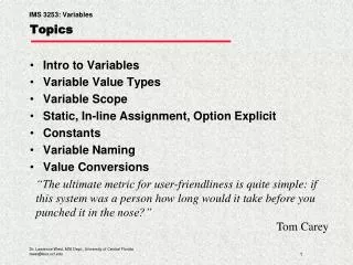
Topics
Topics. Kohler Illumination and conjugation planes Dark field microscopy Phase contrast Differential interface contrast (DIC). Conjugated Planes. Eyepiece. Condensor. Tube Lens. Eye. Objective. Field Diaphgram plan. Intermediate Image plane. Sample plane. Retina. Image Planes.

Topics
E N D
Presentation Transcript
Topics • Kohler Illumination and conjugation planes • Dark field microscopy • Phase contrast • Differential interface contrast (DIC)
Eyepiece Condensor Tube Lens Eye Objective Field Diaphgram plan Intermediate Image plane Sample plane Retina Image Planes
Condensor Tube Lens Objective Field Diaphgram plan Intermediate Image plane Sample plane Image Planes CCD chip
How to avoid image formation -- the opposite of conjugated planes
Eyepiece Collection lens Condensor Tube Lens Eye Objective Field Diaphgram plan Intermediate Image plane Sample plane Retina Back focal plane Aperture Diaphgram plane Lamp Plane
Collection lens Condensor Tube Lens Eye Objective Field Diaphgram plan Sample plane Retina Back focal plane Aperture Diaphgram plane Lamp Plane Retina
Objective Sample plane Back focal plane Close Aperture Smaller Light Cone Angle Smaller NA Less Resolution
Collection lens Condensor Tube Lens Eye Objective Field Diaphgram plan Sample plane Retina Back focal plane Aperture Diaphgram plane Lamp Plane Retina
Objective Condensor Aperture Diaphgram plane Sample plane Back focal plane Close Aperture Smaller Light Cone Angle Smaller NA Less Resolution
Darkfield Microscopy Objective collects Only diffracted light Image plane Objective lens Specimen Condensor allows only high angle rays Stop in condenser Source
Where is light in darkfield? Any refractive index change will scatter light. Scattering redirect light through objective lens.
Comparison of bright and darkfield Brightfield Microscopy Darkfield Microscopy Contrast is reversed in these modalities
Key Requirement of Darkfield: NA of Condendor > NA of Objective
High NA darkfield condenser Visualize particles down to ~40 nm by scattering But not resolvable
Darkfield, Red filter Darkfield Brightfield, High contrast Closed aperture Loss of resolution
Effect of Path Length and Refractive Index Phase shift: Effective Pathlength:
Amplitude vs Phase Objects Amplitude Objects: Cells with stains, pigments, absorb, scatter light, change its amplitude Most biological objects do not absorb white light But diffract, phase shift: phase objects But may be hard to see with contrast by bright field Use phase differences to visualize differences in refractive index: Phase contrast microscopy Nobel Prize in 1953: Zernike
Interferometer: Convert Phase Amplitude Michelson interferometer
Comparison of contrast in brightfield and phase Can see by brightfield, but phase much better
Phase Contrast Microscopy: • Visualize specimens without labeling • Utilizes refractive index for phase differences • Measure thicknesses
Phase Contrast Microscopy Illumination (zero order) Light diffracted by specimen Longer pathway Condenser annulus Condenser Phase rings Objective lens specimen
Phase contrast: Kohler-same Conjugate planes as brightfield Condenser annulus replaces condenser variable diaphragm Objective has phase ring in Backfocal plane S wave projects bright image of annulus onto back aperture (Kohler illumination, conjugate Image planes) Diffracted waves traverse whole back aperture Surround waves un- deviated
Conversion of phase to amplitude Sample retards by λ/4 Phase ring retards by λ/4 (S advanced) S,D shifted by λ/2 Combine and interfere destructively In image plane S is advanced because Of recessed (shorter) path S,D different places in bfp Retarding Plate near Back focal plane
Phase plate located near back focal plane Shaded to attenuate by 70% for more contrast Selectively manipulate either S,D wave components Advances or retards S wave relative to D wave With phase plate
Path Difference between S,D Waves ~ λ/4 For individual cells in tissue culture, the optical path difference relative to fluid is relatively small: A typical cell in monolayer culture: thickness around 5 micrometers refractive index of approximately 1.36. The cell is surrounded by a nutrient medium: refractive index of 1.335 Optical path difference Gives optical path difference of 0.125 micrometer, 125 nm Or λ/4 quarter wavelength (of green light). Subcellular structures (organelles) generate much smaller retardations.
¼ out of phase ½ out of phase PHASE CONTRAST Phase Ring in objective Annulus in condenser destructive interference in phase
2 forms on phase contrast: Positive and Negative Positive: S is advanced higher refractive index= Dimmer image, P is smaller More common Negative: S is retarded higher index= brighter image, P is larger Cells brighter against background Inverse contrast between The 2 modes
Annuli must be perfectly aligned for best contrast Properly aligned Misaligned, blurred
Comparison of phase and DIC microscopy DIC Phase Contrast Phase: path length difference: denser:darker DIC: gradient, not quantitative
DIC is form of Kohler illumination Without Wollastons, reduces to pol scope- Crossed polarizers Also need strain-free objective lenses: Like in pol scope
Selects one polarization Wollaston splits (shears) into perpendicular polarized O,E rays. Located at condensor Front focal plane (Kohler) Shear axis is angle between wedges Recombine in second Wollaston (near Back focal plane) Analyzer in infinity region Between tube lens and objective: Crossed wrt to first polarizer: Cancels action if no change in Refractive index Need coherence for interference in image plane Non-retarded light is blocked by analyzer
Differential Interference Contrast (DIC) Microscopy:uses birefringent prisms to separate light into ordinary, extraordinary components Measure refractive index gradients by relative Retardance of the two beams: no birefringence required in specimen Birefringent Prisms Glan-Thompson Wollaston Used in DIC
From specimen Nomarski is modified: Small offset in wedges Beams combine before: Interference plane needs to be at Back focal (diffraction) Plane. Usually inside lens Nomarski eases alignment Positive Birefringence (quartz): ordinary ray faster, travel through More of the prism, waves “catch up” at exit air interface (regular Wollaston) Calcite has negative birefringence: above argument is opposite O,E Rays differentially retarded when different Optical Path Differences Exist in specimen OPD=
Bias is selective retarding of ordinary ray to increase contrast Symmetric oil droplets Maximum extinction along shear axis Sliding analyzing Wollaston changes phase relationship Of the two beams: change linear plane or ellipticity by path length By changing extinction across field of view: Pseudo 3d relief Bias increases contrast, bright, dark patterns on gray background http://micro.magnet.fsu.edu/primer/java/dic/imagecontrast/index.html
Image Formation in DIC Microscopy Polarizer passes One component Exits prism Elliptically polarized Larger phase change More passed by analyzer More contrast No change in index: No image in a,b (assuming no optical path Difference in two spots) Different paths through Prisms compensate after second
Bias through de Senarmount compensation: quarter wave plate Quarter wave plate, (λ/4) turns linear into circular or elliptical polarization Either between polarizer and condenser prism Or between objective prism and analyzer Then rotate analyzer
Combining linear polarized light OUT OF PHASE IN PHASE Elliptical Polarization Linear Polarization
Circularly Polarized Light • Decompose to linear polarized light with 1/4 phase shift. • No direction (always pass 50% through polarizer in dependent of polarizer orientation) • NOT the same as unpolarized light. • Can be converted back to linear polarized light with birefringent materials (1/4 wave plate).
Principles of Differential Interference Contrast (DIC) DIC turns optical path differences into amplitude contrast DIC is based on retardance introduced by optics-not specimen Different indices used to converts optical path, phase gradients (refractive index) to amplitude differences in the image Recall phase contrast measures actual optical pathlength DIC Not quantitative of actual refractive indices: only gradients Not indicative of actual topography OPD=
Orientation of Object relative to light polarization can give different images, information Perp to shear Parallel to shear Water flea Gill ribs Fish scale Diatoms Orthogonal features can be seen in each case
Thick specimens in Phase and DIC Phase DIC Butterfly wing Tapeworm eggs hydrozoan DIC does better with thicker specimens