
Brain's Protective Coverings and Blood Flow
E N D
Presentation Transcript
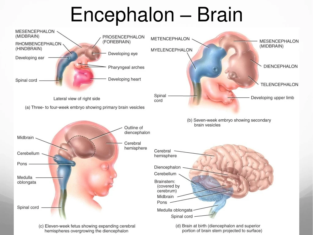
Brain ComponentsFig 14.1 • Brain Stem • Midbrain, Pons, Medulla Oblongata • Cerebellum • “Little Brain” • Diencephalon • Thalamus, Hypothalamus, Epithalamus • Cerebrum
Protective Coverings of the Brain Cranial bones Meninges Dura mater Arachnoid mater Pia mater
Cranial Bones • Cranial vault formed by 8 cranial bones • Encloses and protects brain • Floor divided into 3 fossa: • Anterior – front lobes • Middle – temporal lobes and base of diencephalon • Posterior - cerebellum
Meninges Surround and protect the brain and spinal cord Three membranes: 1. Dura mater 2. Arachnoid mater 3. Pia mater
Dura Mater • Tough outermost meninx. • Composed of two layers: • Periosteal • Meningeal • Venous sinuses between the layers.
Dura Mater • Falx cerebri • Falx cerebelli • Tentorium cerebelli Separates cerebrum from cerebellum Separates 2 hemispheres of cerebrum Separates 2 hemispheres of cerebellum
Arachnoid Mater • Middlemost meninx • Characterized by its filmy, weblike structure • Loosely follows contours of cerebral structures but lies over sulci
Pia Mater • Thin, delicate innermost meninx • Closely adheres to surface of brain and follows sulci and fissures • Provides support for blood vessels serving brain tissue • Sheath of pia mater
Meningeal Spaces • Real or potential spaces between meningeal layers: • Epidural Space = potential space • Between Skull and Dura • Subdural Space = Real space • Between dura and arachnoid • Small bridging veins (little support) cross the space • Subdural hematoma • Subarachnoid space is a real space • Lies between the arachnoid and pia mater • Contains cerebrospinal fluid • Subarachnoid hemorrhage
Brain Injuries • Hemorrhage – active or ongoing bleeding. • Hematoma – accumulation of blood within one of the meningeal spaces or surrounding tissues. • Contusion – Type of hematoma. Blood escapes ruptured capillaries and enters surrounding tissue.
Brain Blood Flow Internal carotid arteries Vertebral arteries Internal jugular veins
Arterial Brain Blood Flow • ANTERIOR: • Common Carotid Arteries Internal Carotid Arteries • Base of skull Cranium • Branch to anterior and middle cerebral arteries
Arterial Brain Blood Flow • POSTERIOR: • Subclavian arteries Vertebral arteries • Transverse foramina of cervical vertebrae foramen magnum • Join at junction of pons and medulla Basilar artery Pontine branches • Posterior inferior cerebellar arteries (off the vertebral artery) • Basilar divides at midbrain anterior inferior cerebellar and superior cerebellar arteries
Arterial Brain Blood Flow • Vertebral arteries • Subclavian • Cervical vertebrae transverse foramina • Basilar artery • Cerebellum, brainstem and occipital lobes. • Pontine arteries • Cerebellar arteries: • Cerebellum • Cerebral arteries • Internal Carotid Artery • Posterior communicating artery • Anterior communicating artery
Arterial Brain Blood Flow • Terminal branches of Carotid and Vertebral arteries = circle of Willis • Circulatory anastomosis (connection between 2 blood vessels) • Backup route in case of blockage • Branch into various cerebral arteries to supply brain with blood
Brain Blood Flow Animation https://www.youtube.com/watch?v=MPcO2ibO75o