Understanding the Cardiovascular System: Heart Function, Structure, and Conduction
220 likes | 335 Vues
The cardiovascular system is crucial for sustaining life, with the heart beating 72 times per minute, totaling around 100,000 beats daily and over 3 trillion beats in a lifetime. It circulates 5-7 liters of blood through arteries and veins, transporting nutrients, oxygen, and waste while regulating hormones and body temperature. The heart consists of four chambers and is structured with specialized tissues such as the endocardium, myocardium, and epicardium. Additionally, the cardiac conduction system ensures coordinated heartbeats through electrical impulses generated by the SA and AV nodes and transmitted via the Bundle of His and Purkinje fibers.

Understanding the Cardiovascular System: Heart Function, Structure, and Conduction
E N D
Presentation Transcript
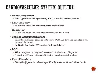
Heart Beats 72 times a minute 100,000 times a day 3 Trillion times in a lifetime! Circulates about 5-7 liters of blood Blood Vessels Arteries Veins Structures
Functions • Transport nutrients and oxygen • Transport waste to kidneys • Distribute hormones and antibodies • Help control body temperature and maintain homeostasis
Heart • 2 Sided double pump • Is about the size of your fist • Lies in the thoracic cavity between the lungs
Heart Tissue • Endocardium: smooth membranous lining inside the heart • Myocardium: thickest layer, muscle tissue that is contractile.
Epicardium: outermost layer in the pericardium Pericardium: covers the outside of the heart Heart Tissue Cont’d
Parts of the Heart • Divided into right and left sides • 2 chambers in each side, for a total of 4 chambers • Atrium: top, where blood enters • Ventricles: bottom, where blood leaves • Left and right sides separated by a partition called a septum
Parts of the Heart Left Atrium Right Atrium Left Ventricle Right Ventricle Septum
Heart Circulation • Pulmonary: Flow of blood between the heart and lungs • Systemic: Flow of blood between the heart and the cells of the body • Coronary: Flow of blood within the heart
Blood flow • Review with heart anatomy
Blood Flow • Vessels • Arteries carry blood away from the heart • Largest = Aorta • Heart muscle contractions pump blood through arteries Veins carry blood towards the heart Largest = Superior/Inferior Vena Cava Valves prevent blood from returning to heart skeletal muscle contractions move blood through veins
Blood Flow Cont’d • Valves • control blood flow • Valve between left atrium and ventricle = bicuspid • Valve between right atrium and ventricle = tricuspid • Pulmonary and aortic valves stop the back flow of blood into the heart
Review Blood flow: • 4 square blood flow color sheet
Cardiac Conduction System • Electrical Impulses produce a wave that can be recorded on the ECG • Consists of • Sinoatrial (SA) node • Atrioventriclular (AV) node • Bundle of His (AV Bundle) • Bundle Branches • Purkinje Fibers (network)
SA NODE • Located in the upper right part of the atrium • Is a natural pacemaker • Fires at a rate of 60 to 100 times per minute • The heartbeat starts in the SA node
AV NODE • Located in the floor of the right atrium • Delays or slows the electrical impulse • Fires at a rate of 40 to 60 time per minute • Can take over if the SA node is not working
Bundle of His • Located next to the AV node • Transfers the electrical impulse from the atria to the ventricles
Bundle Branches • Located along the left and right side of the interventricular septum • Act as pathways or a fork in the road • Impulses in the bundle branch perform the important work of making the heart muscle contract
Purkinje Network • Provide an electrical pathway for each of the cardiac cells • Activate the left and right ventricles simultaneously causing the ventricles to contract