Download
1 / 27
270 likes | 1.09k Vues
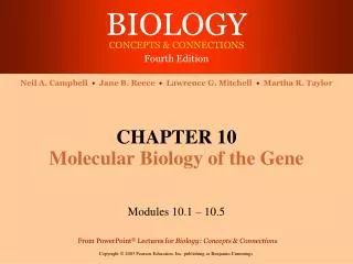
CHAPTER 10 Molecular Biology of the Gene. Modules 10.6 – 10.16. THE FLOW OF GENETIC INFORMATION FROM DNA TO RNA TO PROTEIN. 10.6 The DNA genotype is expressed as proteins, which provide the molecular basis for phenotypic traits.

E N D
CHAPTER 10Molecular Biology of the Gene Modules 10.6 – 10.16
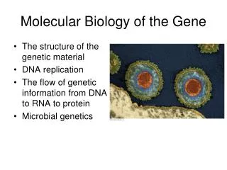
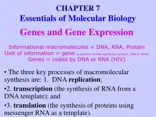
THE FLOW OF GENETIC INFORMATION FROM DNA TO RNA TO PROTEIN 10.6 The DNA genotype is expressed as proteins, which provide the molecular basis for phenotypic traits • The information constituting an organism’s genotype is carried in its sequence of bases
The DNA is transcribed into RNA, which is translated into the polypeptide • A specific gene specifies a polypeptide DNA TRANSCRIPTION RNA TRANSLATION Protein Figure 10.6A
1950’s Protein RNA DNA Phosphorylation Glycosylation Methylation Acetylation Splicing 1980’s DNA RNA Poly- peptide Alternative Splicing Histone modifications MicroRNAs Editing Conformational Isomers Phosphorylation Glycosylation Methylation Acetylation Other Splicing Today DNA RNA Poly- peptide Alternative Splicing Other epigenetic factors Other catalytic regulator RNAs The Evolution of Crick’s Central Dogma from the 1950s to today
10.7 Genetic information written in codons is translated into amino acid sequences • The “words” of the DNA “language” are triplets of bases called codons • The codons in a gene specify the amino acid sequence of a polypeptide
Gene 1 Gene 3 DNA molecule Gene 2 DNA strand TRANSCRIPTION RNA Codon TRANSLATION Polypeptide Amino acid Figure 10.7
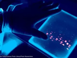
10.8 The genetic code is the Rosetta stone of life • Virtually all organisms share the same genetic code Figure 10.8A
Template strand or antisense - strand Transcribed strand • An exercise in translating the genetic code DNA Coding Strand or Sense + strand Transcription RNA Startcodon Stopcodon Translation Polypeptide Figure 10.8B
10.9 Transcription produces genetic messages in the form of RNA In eukaryotes, RNA poly 1 Synthesizes rRNA, II synthesizes mRNA, and III synthesizes tRNA. RNA poly. Has 5 Subunits: 2 alpha bind reg- ulatory subunits, 1 beta binds the DNA template, 1 beta binds the nucleosides, and one sigma recognizes the promoter and initiates synthesis. RNA nucleotide RNApolymerase Direction oftranscription Templatestrand of DNA Newly made RNA Figure 10.9A
The enzymes of transcription RNA polymerase I is responsible for transcribing RNA that becomes structural components of the ribosome. Pol 1 synthesizes a pre-rRNA 45S, which matures into 28S, 18S and 5.8S rRNAs which will form the major RNA sections of the ribosome. RNA polymerase II transcribes protein-encoding genes, or messenger RNAs, which are the RNAs that get translated into proteins. Also, most snRNA (splicing) and microRNAs (RNAi). This is the most studied type, and due to the high level of control required over transcription a range of transcription factors are required for its binding to promoters. RNA polymerase III transcribes a different structural region of the ribosome (5s), transfer RNAs, which are also involved the translation process, as well as non-protein encoding RNAs.
RNA polymerase DNA of gene • RNA nucleotides line up along one strand of the DNA following the base-pairing rules at the promoter. A regulatory protein binds at -25 binds the TATAAAA box. • This either allows the Polymerase to transcribe or not. Many other protein factors comprise the transcription complex. • 50 nucleotides/sec • 12 bases in the bubble • No proofreading enzymes like DNA • The single-stranded messenger RNA peels away and the DNA strands rejoin after GC hairpin forming region. Promoter DNA Terminator DNA Initiation • In transcription, the DNA helix unzips Elongation Area shownin Figure 10.9A Termination GrowingRNA Completed RNA http://www.johnkyrk.com/DNAtranscription.html RNApolymerase Figure 10.9B
10.10 Eukaryotic RNA (hnRNA) is processed before leaving the nucleus Exon Intron Exon Intron Exon DNA TranscriptionAddition of cap and tail • Noncoding segments called introns are spliced out • A cap and a tail are added to the ends • 5” cap is a guanosine nucleotide connected to the mRNA via an unusual 5' to 5' triphosphate linkage. This guanosine is methylated on the 7' position directly after capping in vivo by a methyl transferase. • The addition of adenine nucleotides to the 3′ end of messenger ribonucleic acid molecules during posttranscriptional modification Cap RNAtranscriptwith capand tail Introns removed Tail Exons spliced together mRNA Coding sequence NUCLEUS CYTOPLASM Figure 10.10
10.11 Transfer RNA molecules serve as interpreters during translation Amino acid attachment site • In the cytoplasm, a ribosome attaches to the mRNA and translates its message into a polypeptide • The process is aided by transfer RNAs Hydrogen bond RNA polynucleotide chain Anticodon Figure 10.11A
Each tRNA molecule has a triplet anticodon on one end and an amino acid attachment site on the other Amino acidattachment site Anticodon Figure 10.11B, C
10.12 Ribosomes build polypeptides Next amino acidto be added topolypeptide Growingpolypeptide tRNA molecules P site A site Growingpolypeptide Largesubunit tRNA P A mRNA mRNAbindingsite Codons mRNA Smallsubunit Figure 10.12A-C
10.13 An initiation codon marks the start of an mRNA message Start of genetic message End Figure 10.13A
mRNA, a specific tRNA, and the ribosome subunits assemble during initiation Largeribosomalsubunit Initiator tRNA P site A site Startcodon Small ribosomalsubunit mRNA 1 2 Figure 10.13B
10.14 Elongation adds amino acids to the polypeptide chain until a stop codon terminates translation • The mRNA moves a codon at a time relative to the ribosome • A tRNA pairs with each codon, adding an amino acid to the growing polypeptide
Amino acid Polypeptide Asite P site Anticodon mRNA 1 Codon recognition mRNAmovement Stopcodon Newpeptidebond 2 Peptide bond formation 3 Translocation Figure 10.14
10.15 Review: The flow of genetic information in the cell is DNARNAprotein • The sequence of codons in DNA spells out the primary structure of a polypeptide • Polypeptides form proteins that cells and organisms use
TRANSCRIPTION DNA Stage mRNA istranscribed from aDNA template. 1 mRNA RNApolymerase • Summary of transcription and translation Amino acid TRANSLATION Stage Each amino acid attaches to its proper tRNA with the help of a specific enzyme and ATP. 2 Enzyme tRNA Initiator tRNA Anticodon Stage Initiation of polypeptide synthesis 3 Largeribosomalsubunit The mRNA, the first tRNA, and the ribosomal subunits come together. Start Codon Smallribosomalsubunit mRNA Figure 10.15
Newpeptidebondforming Growing polypeptide Stage Elongation 4 A succession of tRNAs add their amino acids to the polypeptide chain as the mRNA is moved through the ribosome, one codon at a time. Codons mRNA Polypeptide Stage Termination 5 The ribosome recognizes a stop codon. The poly-peptide is terminated and released. Stop Codon Figure 10.15 (continued)
10.16 Mutations can change the meaning of genes • Mutations are changes in the DNA base sequence • These are caused by errors in DNA replication or by mutagens • The change of a single DNA nucleotide causes sickle-cell disease
Normal hemoglobin DNA Mutant hemoglobin DNA mRNA mRNA Normal hemoglobin Sickle-cell hemoglobin Glu Val Figure 10.16A
Missense-mutation causing a change in aa. Nonsense-mutation causing a premature stop codon • Types of mutations mRNA NORMAL GENE Protein Met Phe Gly Ala Lys BASE SUBSTITUTION Met Lys Phe Ser Ala Causes a “frame shift” Missing BASE DELETION Met Lys Leu Ala His Figure 10.16B