SQUINT
SQUINT. Dr. Ajay Dudani, Mumbai Retina Centre. DEFINITION.

SQUINT
E N D
Presentation Transcript
SQUINT Dr. Ajay Dudani, Mumbai Retina Centre
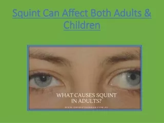
DEFINITION • Squint is a disorder in which one eye misaligns with the other when focusing in a primary direction of gaze. It is an imbalance in the normal tone or coordination of one or more extra ocular muscle which results in a manifest deviation of the affected eye.
CLASSIFICATION 1. Direction of deviation: - convergent (esotropia) - divergent (exotropia) - hypodeviation - hyperdeviation 2. Comitancy: - comitant or non paralytic - incomitant or paralytic
3. Constancy: - intermittent - constant 4. Onset: - childhood (congenital) - adult (acquired) 5. Unilateral or Alternating
6. Apparent (psuedostrabismus) Manifest (tropias) Latent (phorias)
Pseudoesotropia- in prominent epicanthal folds, high myopia Pseudoexotropia- in hypertelorism PSEUDOSTRABISMUS
ETIOLOGY • High Refractive errors - High degree of uncorrected refractive error in children may cause deviation of the most affected eye. • Ocular conditions- causing visual axis obstruction eg. cataract, corneal opacities, retinoblastoma, ROP, macular disease etc.
3. Trauma 4. Lesions affecting the EOM’s or CN’s especially no. III 5. Systemic dis- DM, stroke, botulism
PREDISPOSING FACTORS • General debility and lowered vitality • Psychosis, neurosis and mental stress • Inadequacy of fusional reserve • Precision of job • Advancing age
CAUSES OF CHILDHOOD STRABISMUS • Cataract • Corneal opacities • ROP • Retinoblastoma • Traumatic brain injury • Hemangioma near the eye during infancy • Trisomy 18 • Congenital rubella • Cerebral palsy
SYMPTOMS • Deviated eye • Abnormal head posture • Poor vision • Headache • Diplopia
EXAMINATION 1.HISTORY A. Deviation: Age of onset Description of deviation Previous treatment B. Pre and post natal factors Growth and development Family history of strabismus 2.GENERAL OBSERVATION. Abnormal head posture
3. VISUAL ACUITY a. Without glasses and with glasses b. Near and distant vision c. Amblyopia testing 4.MOTOR: a. Extra ocular movements. b. Phorias or tropias c. Near point of convergence and near point of accommodation.
5. MEASUREMENT OF DEVIATION. Distance and near Without glasses and with glasses( if worn) 6. SENSORY TESTS Worth 4 dot test. Stereopsis 7. FIXATION: monocular , alternating, binocular 8. SLIT LAMP EXAMINATION.
9 . FUNDUS EXAMINATION. 10 . CYCLOPLEGIC REFRACTION.
VISION TESTS • In infants: - fixation and following light - Catford drum test - preferential looking test - Cardiff acuity test - VER - reflex responses
In 1 to 2 yr old: - Boeck candy test - Worth’s ivory ball test • In 2 to 3 yr old: - coin test - miniature toys test - dot visual acquity test
In 3 to 5 yr old: - tumbling E test - Landolt’s C test - Sheridan letter test
SENSORY TESTS • Worth’s 4 dot test • Bielchowsky’s after image test • Striated glasses of Bagolini • 4 diopter prism base out test • Synaptophore
STEREOPSIS TESTS • Titmus stereo test • Random dot stereogram test • Random dot e test • TNO test • Lang test • Frisby test • 2 pencil test
HEAD POSTURE • Incomitant squint • Position of head in which the eyes are in a position of no deviation or very small deviation so that fusion is possible. • 3 components: -Chin -Face turn -Head tilt
TESTS TO MEASURE DEVIATION ANGLE • Hirschberg corneal reflex test • Krimsky’s test • Cover test • Alternate cover uncover test • Prism bar cover test • Maddox wing test • Maddox rod test
MOTILITY TESTS • Ocular movements - versions - ductions • Near point of convergence- RAF rule • Near point of accomodation- RAF rule • Fusional amplitudes- with prism bar or synaptophore
DIPLOPIA TESTS • Hess test • Lees screen
AMBLYOPIA • Unilateral or bilateral DOV due to form deprivation &/or abnormal binocular interaction for which there is no ocular or visual pathway pathology • Most commonly due to squint, large uncorrected refractive errors etc. • Treatment: - occlusion - penalisation
MANAGEMENT • Refractive error correction by spectacle lenses, prisms, orthoptic exercises. • Patching, reducing the VA or occluding the good eye so as to activate the deviated eye. This is only done in children with unilateral deviation.
Medical : - miotics in accomodative esotropia - cycloplegics in accomodative esotropia - levodopa, carbidopa in amblyopia - botulinum toxin A (botox)
Surgery: - weakening procedures eg. recession, marginal myotomy, myectomy. - strengthening procedures eg. resection, tucking, advancement.