Download
1 / 12
120 likes | 303 Vues
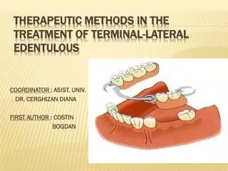
COORDINATOR : ASIST. UNIV. DR. CERGHIZAN DIANA FIRST AUTHOR : COSTIN BOGDAN. THERAPEUTIC METHODS IN THE TREATMENT OF TERMINAL-LATERAL EDENTULOUS.

E N D
COORDINATOR : ASIST. UNIV. DR. CERGHIZAN DIANA FIRST AUTHOR : COSTIN BOGDAN THERAPEUTIC METHODS IN THE TREATMENT OF TERMINAL-LATERAL EDENTULOUS
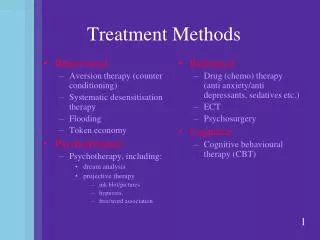
Partiallyedentulous dentitions represents a pathophysiological state of the orofacial system, characterized by the absence of one or more, but not all natural teeth. Common condition among patients with influences upon the quality of their life, with modern therapeutic options. Most commonly used classifications are those developed by Costa and Kennedy. introduction
- 54 years old female patient with a class II, modification 1 Kennedy partially edentulousmaxillary arch, partially treated
exooral EXAMINATIONS • Inspection : symmetric, oval, convex profile, no tegument color changes, vertical dimension of occlusion with no pathologic changes. • Palpation : normal sensitivity and local ganglions without any changes. Higher contraction ratio of the masseter and temporal muscles on the left side.
ENDOORAL EXAMINATIONS • Normal gums, frontal remaining teeth with fillings, color changes, with no pathologic mobility, presenting a dental bridge from 2.3 to 2.8. • Distal residual ridge horizontally with Class I maxillary tuberosity, favorable palate, and a second degree of atrophy -> higher atrophy on the vestibular side because of the centripetal atrophy of the maxillary bone.
OCCLUSION EXAMINATION Sagittal : physiologic occlusion on the canine and incisive Transverse : normal ratio Vertical : normal as well with the exception of the non existing overbite.
After all the treatments that precedes the treatment plan for the removable partial denture were fulfilled the diagnosis of class II, modification 1 Kennedy partially edentulous arch remains unchanged. DIAGNOSIS
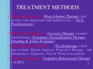
Dental bridge Acrylic removable partial denture Removable partial denture Oral implants Therapeutic options
After visual examination, radiographs, oral prophylaxis, evaluation of teeth and periodontium, vitality tests of individual teeth, and impressions of each arch the most adequate treatment was chosen reporting it to the oral condition, the age, and the financial state.
Chosen treatment In this case we opted for the removable partial denture .
Results • Rehabilitation of occlusion • Higher masticatory efficiency • Bilateral mastication • Improved phonetic • Improvement of aesthetic
VA MULTUMIM !!!