Download
1 / 1
10 likes | 103 Vues
Fall, 2011. Final Semester Project. Biol 443, Biology and Biochemistry of Proteins. A Profile of the Mitto_carr Family. Kirsten West . INTRODUCTION. STRUCTURE. ACTIVE SITE. FAMILY STRUCTURE.

E N D
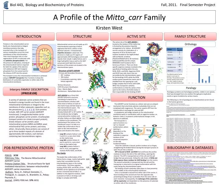
Fall, 2011. Final Semester Project Biol 443, Biology and Biochemistry of Proteins A Profile of the Mitto_carr Family Kirsten West INTRODUCTION STRUCTURE ACTIVE SITE FAMILY STRUCTURE The active site of the ADT1-BOVIN is located at the bottom of the cavity that is formed by the proteins tripartite arrangement of α- helices. All ADP/ATP carriers consist of a triplication of a sequence of 20 to 30 amino acid residues located on the highly kinked odd numbered α-helices (PxD/ExxK/RxK/R-(20 to 30 residues)-D/EGxxxxaK/RG) and the unique RRRMMM motif located at the C terminus of helix 5. There are three specific argenine residues involved in the specificity of binding within the site, R234, R235, and R236. Argenines R234 and R235 have side chains that are accessible by the intermembrane space for the binding of ATP, while the R236 residue points towards the matrix and is shielded from the opening by a salt bridge complex that it forms with E264. When the transporter switches conformations so that the cavity is then open to the matrix, the R236 residue is then responsible for the binding of ADP. Orthology Proteins in the mitochondrial carrier family are characterized as integral membrane proteins that play essential roles in the transport of various metabolites across the inner membranes of the mitochondrion. Their most important role is in the regeneration of ATP in eukaryotic cells via oxidative phosphorylation. For this process to take place, crossing of the mitochondrial matrix is required for both the uptake of the electron rich substances ADP and inorganic phosphate from the cytosol and for the release of ATP into the cytosol. Mitochondrial carriers are all made up of 6 transmembrane spanning a-helical segments that form a dimer or two identical subunits. A characteristic tripartite structure is observed. This structure is made up of 3 regions of approximately 100 homologous amino acid repeats that include 2 alpha helices and 1 large hydrophilic loop as well as the MCF conserved motif. Both the N and C terminus of the protein are located in the intermembrane space. Argenine residue R234, responsible for ATP binding ADP/ATP transporter • Structure of ADT1-BOVIN • Primary and Secondary Structures: • 297 residues • 70% helical • 6 membrane-spanning alpha helical segments Argenine residue R236, responsible for ADP binding. Salt bridge formed with E264 The Mitochondrial Carrier protein is not seen in Archaea. The protein is scarce among bacteria and viruses but seen readily in plants, fungi, and especially animals This transport across the mitochondrial membrane is achieved using an ADP/ATP transporter that functions to keep the distribution of nucleotides equimolar on either side of the mitochondrial membrane. Tertiary Structure: CATH Structure Classification Class: Mainly Alpha Architecture: Alpha/Alpha Barrel ADT1-BOVIN has a three-fold pseudo-symmetric structure, consisting of a barrel of six transmembrane α-helices with three short α-helices on each of the matrix loops and two loops in the intermembrane space . The odd numbered helices are sharply kinked (20oto 35o) within the membrane due to highly conserved proline residues and all other helices are tilted slightly in relation to the membrane. The positioning of the helices in the membrane cause it to take on a basket-like conformation that is open to the intermembrane space and closed to the matrix. Paralogy Interpro FAMILY DESCRIPTION (IPR018108) Paralogous proteins are homologous proteins, visible in one species, that have diverged from one another through gene duplication events or gene mutations by various means. The following is a list of paralogues to mitochondrial carriers found in the human genome: FUNCTION A variety of substrate carrier proteins that are involved in energy transfer are found in the inner mitochondrial membrane or integral to the membrane of other eukaryotic organelles such as the peroxisome [1, 2, 3, 4, 5, 6]. Such proteins include: ADP, ATP carrier protein (ADP/ATP translocase); 2-oxoglutarate/malate carrier protein; phosphate carrier protein; tricarboxylate transport protein (or citrate transport protein); Graves disease carrier protein; yeast mitochondrial proteins MRS3 and MRS4; yeast mitochondrial FAD carrier protein; and many others. Structurally, these proteins can consist of up to three tandem repeats of a domain of approximately 100 residues, each domain containing two transmembrane regions. The ADP/ATP carrier functions as a dimer and uses an antiport mechanism that only binds adenine nucleotides that are not complexed with magnesium. The carrier exports ATP from the mitochondria in exchange for external ADP. A cytoplasmic salt bridge network on the mitochondrial membrane works in coordination with an argenine residue, on kinked α- helix 5, to prevent conformational changes in the absence of substrate. Database: NCBI Protein Reference Sequences 11,008,086 sequences; 3,852,209,289 total letters Query= >2C3E:A|PDBID|CHAIN|SEQUENCESDQALSFLKDFLAGGVAAAISKTAVAPIERVKLLLQVQHASKQISAEKQYKGIIDCVVRIPKEQGFLSFWRGNLANVIRYFPTQALNFAFKDKYKQIFLGGVDRHKQFWRYFAGNLASGGAAGATSLCFVYPLDFARTRLAADVGKGAAQREFTGLGNCITKIFKSDGLRGLYQGFNVSVQGIIIYRAAYFGVYDTAKGMLPDPKNVHIIVSWMIAQTVTAVAGLVSYPFDTVRRRMMMQSGRKGADIMYTGTVDCWRKIAKDEGPKAFFKGAWSNVLRGMGGAFVLVLYDEIKKFV Length=297 Sequences producing significant alignments: Score(Bits) E Value ref|NP_001142.2| ADP/ATP translocase 1 [Homo sapiens] 585 0.0 ref|NP_001627.2| ADP/ATP translocase 3 [Homo sapiens] 558 0.0 ref|NP_001143.2| ADP/ATP translocase 2 [Homo sapiens] 557 0.0 ref|NP_112581.1| ADP/ATP translocase 4 [Homo sapiens] 444 2e-161 ref|NP_037518.3| calcium-binding mitochondrial carrier protei... 130 2e-37 ref|NP_998816.1| calcium-binding mitochondrial carrier protei... 130 2e-37 ref|NP_689920.1| graves disease carrier protein [Homo sapiens] 118 7e-34 ref|NP_077008.2| calcium-binding mitochondrial carrier protei... 117 2e-32 ref|NP_003696.2| calcium-binding mitochondrial carrier protei... 115 3e-31 ref|NP_848621.2| solute carrier family 25 member 42 [Homo sap... 111 3e-31 ref|NP_055066.1| calcium-binding mitochondrial carrier protei... 112 2e-30 ref|NP_001153682.1| calcium-binding mitochondrial carrier pro... 112 2e-30 ref|NP_001006644.1| calcium-binding mitochondrial carrier pro... 109 2e-30 ref|NP_775908.2| solute carrier family 25 member 41 [Homo sap... 108 8e-30 ref|NP_660348.2| solute carrier family 25 member 43 [Homo sap... 107 9e-30 ref|NP_001006642.1| calcium-binding mitochondrial carrier pro... 109 1e-29 ref|NP_443133.2| calcium-binding mitochondrial carrier protei... 108 2e-29 ref|NP_001006643.1| calcium-binding mitochondrial carrier pro... 108 2e-29 ref|NP_113669.1| mitochondrial glutamate carrier 2 [Homo sapi... 104 8e-29 ref|NP_001010875.1| kidney mitochondrial carrier protein 1 [H... 104 8e-29 The transporter is said to function in a ‘pit’ to ‘channel’ conformation, where opening is dependent on the binding of the substrate to argenineresidues of the conserved sequence on the odd numbered helices. • Loop MI contains helices I and II and h1-2, a very short helix on the matrix loop • Loop MII contains helices III and IV and h3-4, a very short helix on the matrix loop • Loop MIII contains helices V and VI, h5-6, and argenine residues R234, R235, and R236 BIBLIOGRAPHY & DATABASES PDB REPRESENTATIVE PROTEIN Once the substrate is bound, prolineresidues act as hinges, straightening the kinked helices during the transport process to open the pit into a channel. ATP is bound to R234 and R235 first and forces the proline residues to loosen the kink of the odd helices, opening the channel. The ATP is transported out of the cell and the salt bridge that was previously hiding the side chains of R236 and E264 exposes these residues for ADP bindingfrom the matrix. The second nucleotide is transported into the cell and the transporter is returned to its original conformation with the cavity facing the intermembrane space. The binding of both substrates takes place in close proximity to the middle of the mitochondrial membrane. Once a nucleotide is bound from either side of the carrier, it favors binding of the second one from the opposite side, preventing binding of two nucleotides from the same side on each monomer. Consequently, the exchange of the substrates will be equimolarand dependent on the presence of substrate on either side of the membrane. • BLASTP 2.2.26+ Reference: Stephen F. Altschul, Thomas L. Madden, Alejandro A. Schaffer, Jinghui Zhang, Zheng Zhang, Webb Miller, and David J. Lipman (1997), "Gapped BLAST and PSI-BLAST: a new generation of protein database search programs", Nucleic Acids Res. 25:3389-3402. • The Pfam protein families database: R.D. Finn, J. Mistry, J. Tate, P. Coggill, A. Heger, J.E. Pollington, O.L. Gavin, P. Gunesekaran, G. Ceric, K. Forslund, L. Holm, E.L. Sonnhammer, S.R. Eddy, A. BatemanNucleic Acids Research (2010) Database Issue 38:D211-222 • http://www.ebi.ac.uk/interpro/DisplayIproEntry?ac=IPR018108 • Edmund R.S Kunji, The role and structure of mitochondrial carriers, FEBS Letters, Volume 564, Issue 3, 30 April 2004, Pages 239-244, ISSN 0014-5793, 10.1016/S0014-5793(04)00242-X.(http://www.sciencedirect.com/science/article/pii/S001457930400242X)Keywords: Mitochondrial carrier; Electron crystallography; X-ray crystallography; Structure; Membrane protein • F. Palmieri, Mitochondrial carrier proteins, FEBS Letters, Volume 346, Issue 1, 6 June 1994, Pages 48-54, ISSN 0014-5793, 10.1016/0014-5793(94)00329-7.(http://www.sciencedirect.com/science/article/pii/0014579394003297)Keywords: Carrier protein; Transmembrane topology; Bacterial expression; Transport; Liposome; Mitochondrion • John E. Walker, The mitochondrial transporter family, Current Opinion in Structural Biology, Volume 2, Issue 4, August 1992, Pages 519-526, ISSN 0959-440X, 10.1016/0959-440X(92)90081-H.(http://www.sciencedirect.com/science/article/pii/0959440X9290081H) • Lucy R. Forrest, ReinhardKrämer, Christine Ziegler, The structural basis of secondary active transport mechanisms, Biochimica et BiophysicaActa (BBA) - Bioenergetics, Volume 1807, Issue 2, February 2011, Pages 167-188, ISSN 0005-2728, 10.1016/j.bbabio.2010.10.014.(http://www.sciencedirect.com/science/article/pii/S000527281000722X)Keywords: Secondary active transport; Protein structure; Carrier; Coupling; Modeling; Protein conformation • Title: Relations between structure and function of the mitochondrial ADP/ATP carrier Author(s): Nury H. ; Dahout-Gonzalez C. ; Trezeguet V. ; et al.Source: ANNUAL REVIEW OF BIOCHEMISTRY Book Series: Annual Review of Biochemistry • Biophysics: Yi Wang and EmadTajkhorshid; Electrostatic funneling of substrate in mitochondrial inner membrane carriersPNAS 2008 ; published ahead of print July 8, 2008,doi:10.1073/pnas.0801786105 The Cavity The basket like conformation of the helices forms a large, mainly hydrophilic cavity that is accessible from the intermembrane space in the resting conformation. The three argenine residues R234, R235, and R236, of the ADP/ATP carrier signature are all located on the C-terminal end of helix 5. In addition to the argenine residues, E264 is present on helix 5, forming a salt bridge in complex with R236. PDB ID:2C3E PDB Entry Title: The Bovine Mitochondrial ADP/ATP Carrier Primary Citation Title: Structural basis for lipid mediated interactions between mitochondrial ADP/ATP carrier monomers Authors:Nury, H., Dahout-Gonzalez, C., Trezeguet, V., Lauquin, G., Brandolin, G., Pebay-Peyroula, E., Journal:(2005) FEBS lett. 579: 6031