Download
1 / 18
210 likes | 1.98k Vues
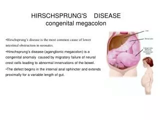
HIRSCHSPRUNG'S DISEASE congenital megacolon. Hirschsprung’s disease is the most common cause of lower intestinal obstruction in neonates.

E N D
HIRSCHSPRUNG'S DISEASEcongenital megacolon Hirschsprung’s disease is the most common cause of lower intestinal obstruction in neonates. Hirschsprung’s disease (aganglionic megacolon) is a congenital anomaly caused by migratory failure of neural crest cells leading to abnormal innervations of the bowel. The defect begins in the internal anal sphincter and extends proximally for a variable length of gut.
Incidence & Etiology • INCIDENCE: 1\5000 live birth newborn • 70-80% is boys. (M / F. 4: 1 ) • Less common in blacks.
PATHOPHYSIOLOGY EMBRYOLOGY: • The neuroenteric ganglion cells migrate from the neural crest to the upper end of the alimentary tract and then follow the vagal fibers caudally • delay or arrest in this migration results in the neural crest cells failing to reach the distal bowel
PATHOPHYSIOLOGY • The fundamental pathology in HD is the absence of ganglion cells in the submucosal and intermuscular nerve plexuses and is associated with an increase in the nerve fibers in the affected segment . • That aganglionic segment usually involves the terminal intestine, i.e. the rectum or rectosigmoid. The aganglionic segment may, however, include the entire large bowel and even small bowel. • The gross pathologic feature of HD is a dilated proximal intestine with gradual or abrupt transition to normal calibrated distal intestine . The TZ is typically funnel like or cone shaped . • The colon proximal to the aganglionic segment, in an effort to overcome the partial obstruction, becomes distended and its wall markedly thickened because of muscle hypertrophy • The degree of hypertrophy and dilatation depends upon the duration and degree of obstruction and thus, indirectly to the age of the patient.
TYPES. • Congenital : This type is the commonest one . • Etiology of the disease is still unknown.but Genetic factors are now identified. • 10% of cases have familial history, especially those with long segment disease. • Acquired : Degeneration of the ganglions may occur due to: -Vascular causes like after pullthrough procedure due to ischemia & tension. - Non vascularcauses like Trypanosoma (chaga's disease). Vit B1 def. Chronic infection ( TB.).
ASSOCIATED ANOMALIES • HD is usually a solitary anomaly in a full term, otherwise healthy infant • Associated anomalies do occur in nearly 20% of cases • urogenital system (11%) • cardiovascular system (6%) • gastrointestinal system (6%), • with 8% having various other malformations • Prematurity is reported in as many as 10% of those children with HD • Trisomy 21 occurs in approximately 5% of cases
CLINICAL PRESENTATIONS : • Failure to pass meconium in the 1st 24h of life 98% of neonates pass meconium in the first 24 hours of age.. Any newborn who fails to pass meconium in the first 24-48 hours of life should be evaluated for possible Hirschsprung's disease. • Neonatal Intestinal obstruction symptoms include bilious vomiting, abdominal distension and refusal to feed. • Recurrent Enterocolitis mainly in the 1st three months of life. • TOXIC MEGACOLON : Fever. Abdominal distension. Bile stained vomitous. Explosive diarrhoea. Dehydration. Shock. • Spontanous perforation occurs in 3%,specially if long segment aganglionosis. • Chronic constipation patients may have chronic constipation in response to changes in feeding. And may have Growth retardation. Multiple fecal masses on abdominal examination.
Diagnosis History failure to pass meconium, painless abdomenal distension & constipation) Physical examinations Distended abdomen with Multiple fecal masses on abdominal examination on DREcharacteristically there is • Anal sphincter is hypertonic • Rectum is typically empty. • Hard fecal mass. Radiology • Plain x-rays of the abdomen :Erect & supine • Contrast Enema. Shows narrow distal segment,funnel-shaped dilatation at level of transition zone with marked dilatation of the proximal colon. 24-hrs delayed films is important in diagnosis; it shows poor emptying with barium throughout the colon, as opposed to the child with psychogenic stool holding in whom the barium generally collects in the distal rectosigmoid. contrast enema should be done with out preparation of bowel,
Electromanometry : • not useful in neonate • excellent screening tool in infant & children . • The classic finding is the absence of the recto anal inhibitory reflex when the rectum is distended. • Rectal biopsy : • Rectal biopsy is the definitive diagnostic test and demonstrates absence of ganglion cells, nerve hypertrophy and stains indicating increased acetylcholinesterase activity. • suction mucosal biopsy (at different levels ). Can be done without anesthesia • full thickness biopsy is done under general anesthesia. • UltraSonography: for associated anomalies.
Management : Manegement of HD differs accosrding to the presentation form and clinical situation of the patients: • Acute I.O. : if the patient presents with acute intestinal obstruction in the early life the management will be • resuscitation , • NGT , NPO • IVF , • Antibiotics , • Rectal tube,irrigations . • The initial treatment requires performing a "leveling" colostomy in the most distal colon with ganglion cells present. This requires exploration with multiple seromuscular biopsies of the colon wall to determine the exact extend of the aganglionosis. The colostomy is placed above the transition zone. Placement of the colostomy in an area of aganglionosis will lead to persistent obstruction • When the patient becomes stable, then the definitive treatment will be planned. • Chronic constipation : • laxative • saline enema. • Work up to establish the diagnosis • then the definitive treatment will be planned
Definitive procedures: Once the child has reached an adequate size and age (6-12 months; 20 pounds or more), a formal pull-through procedure is done • Open surgery : There are many surgical options for Pull-through operation.All aiming at resection of aganglionic segment and anastomosing the two normal ganglionic ends.They give excellent result in 90%. a.swenson. b.soave. c.Rehbein. d. Duhamel. e. Boley's. • LAPAROSCOPY . • TEPT transanal endorectal pullthrough (without laparotomy )
COMPLICATIONS of Pullthrough • anastomotic leak. • stricture . • retraction of the colon. • fecal incontinence (soiling or encopresis ). • persistant constipation.
Distinguishing features between childhood functionalconstipation and Hirschsprung’s disease
Back Fig. 1 Plain X-ray showing dilated loops of bowel with absence of gas in the rectum
back TZ
Back Fig. 2 Contrast Enema showing Aganglionic segment with the transition zone