Download

1 / 22
220 likes | 346 Vues
Learn about the functions and structures of the endocrine system, including the pituitary gland, thyroid gland, parathyroid glands, and adrenal glands. Explore hormone release mechanisms and glandular components in detail with anatomical descriptions.

E N D
Mechanisms of hormone release (a) Humoral: in response to changing levels of ions or nutrients in the blood (b) Neural: stimulation by nerves (c) Hormonal: stimulation received from other hormones
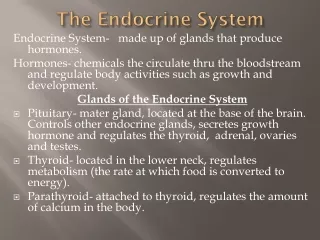
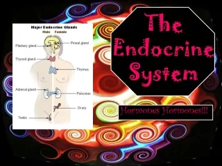
1.Hypophysis (Pituitary Gland) • The hypophysis consists of glandular(Adenohypophysis) & nervous portions(Neurohypophysis) At low power, identify: • Adenohypophysis: • Pars anterior (pars distalis; anterior pituitary). • Pars intermedia( remnant of rathke pouch rudimentary in human beings) . • Pars tuberalis • Neurohypophysis: • Pars nervosa ( posterior pituitary) consist of median eminence and infundibulum.
Anterior Pituitary (Pars Distalis /Pars Anterior) • Cells can be divided into two classes on the basis of their staining characteristics: • Chromophils ("color-loving"), and chromophobes("color-fearing"). • Chromophils are further classified as acidophils and basophils. • Each cell type produces only one type of hormone. • parenchyma consists of secretory epithelial cells arranged in anastomosing cords and clusters. • Abundant sinusoidal capillaries with fenestration
Anterior pituitory… Chromophil: • Acidophils ; • strongly-staining, acidophiliccytoplasm due to granules. • cells are larger,most abundant than chromophobes. • well developed cell outline • Basophils- • Cells are variable in sixe and shape with basophilic cytoplasm. • secretory granules responsible for the staining characteristics of these cells are.
Anterior pituitory… Chromophobe- • clear, faintly-staining, sparse cytoplasm. • Do not have granules • Indistinct cell outline • Represent chromophil without granules or stem cells • Sinusoidal Capillaries in Anterior Pituitary • Chromophil granules are dense cored vesicles
Pars Intermedia… • Intermediate portion of pituitary • part of the adenohypophysis, • is non-neural. • composed of a thin layer of epithelial cells, which enclose colloid-filled spaces. • Identify colloid vesicles, and the surrounding low columnar cells .
Pars Nervosa… • is a downgrowth from the hypothalamus • Exhibits characteristics of nervous tissue. • Axon terminals within this area originate from cells in the hypothalamus; those in the supraoptic nucleus produce ADH (vasopressin), while those in the paraventricular nuclei produce oxytocin. • Cell bodies of these neurons are located in the hypothalamus, • The pars nervosa contains no neuronal cell bodies.
Pars nervosa… • Pituicytes : • Glial cells, variable in cell size and shape. • barely visible having orange stained cytoplasm. • Herring bodies: • large unmyelinated axon terminals, containing large numbers of neurosecretory granules. • Rich capillary network present in pars nervosa
Thyroid Gland-General Structure • It stores the inactive form of hormone extracellularly in follicles . • General Structure. Identify … • connective tissue capsule . • connective tissue septa that divide the bilobed gland into lobules .
follicles -of various sizes, • filled with pink-stained colloid and lined by cuboidal epithelial cells. These are the • functional units of the thyroid gland. • follicle size varies inversely with secretory activity. Interfollicular regions – • the presence of connective tissue, sinusoidal capillaries into which hormone is released) • parafollicular cells
Thyroid Follicles • Follicular epithelium: • vary from high cuboidal to low cuboidal ; reflects the level of follicle secretory activity. • follicle cells (principal cells) have large, centrally- or basally-located nuclei, • cells active in protein secretion. • Storage and release of thyroid hormones involves the protein thyroglobulin .
Parafollicular Cells • Found scattered singly or in small groups present in periphery of the follicles , • cells are responsible for production of calcitonin, a peptide hormone that is synthesized and secreted independently of thyroid hormone. • Also called C, clear, or light cells.
Parathyroid Glands-Parenchyma • present as a mass of crowded single cells, not arranged as follicles. It consist of 2 type of cells: Chief cells (principal cells): • Numerous ,small cells withprominent nuclei, and the pale,scant cytoplasmic staining.
Parathyroid Glands… Oxyphil cells: • single or clumps of larger cells with acidophilic (oxyphilic), cytoplasm with dark staining nucleus. • Number inrease with age. • Oxyphil cells are not always present in a section of parathyroid gland.
Adrenal Glands… Like the pituitary, they are composed of two distinctly different components, one of mesothelial origin and one of neural origin . General Structure • outer cortex , and the inner medulla . These zones are readily observed even in a fresh, unstained section. • 3 histological zones of the cortex. • tough connective tissue capsule and radial trabeculae that extend into the cortex. • Prominent central veinin the medulla.
Adrenal Cortex: Zona Glomerulosa Identify… • outermost cortical layer , and note the presence of columnar epithelial cells arranged in long cords that appear as ovoid clumps when cut in cross-section. • cytoplasm is pink and relatively scant contain lipid droplets. • capillary sinusoids are abundant.
Adrenal Cortex: Zona Fasciculata… • Note that this is the broadest, lightest-stainingof the three cortical zones, with epithelial cells that have large, abundant, poorly-stained cytoplasm. • Cells are arranged in vertical columns of radial plates • Identify capillary sinusoids between cords of secretory cells.
Adrenal Cortex: Zona Reticularis… • Note that the epithelial cells of this innermost, prominently stained zone are arranged in irregular, anastomosing cords and clumps with wide capillary sinuses intervening. • Note that the secretory cells are small, with relatively darkly stained cytoplasm that may contain yellow pigment.
Adrenal Medulla: Chromaffin Cells • Derived from neural crest, cells of the medulla are functionally equivalent to postganglionic sympathetic neurons. • Chromaffin cells. The secretory cells of the medulla contain catecholamines (norepinephrine and epinephrine) in cytoplasmic granules that are oxidized to a brown color by potassium bichromate (the chromaffin reaction).
Adrenal Medulla: Chromaffin Cells • Note that chromaffin cell cytoplasm is quite basophilic, compared to the acidophilia of the adjacent zona reticularis. • Note that the cells are arranged into tight clumps, with wide capillaries and venous channels intervening .
Pineal Gland-Pineal Sand • Basophilic, extracellular concretions . They are often calcified, which makes the pineal gland an excellent radiological marker, particularly of the midline.