Schematic Diagrams of EGFR and RIP1 Promoters and Their Mutants Linked to Luciferase
This illustration presents a detailed schematic diagram comparing full-length and truncated EGFR promoters linked to the firefly luciferase gene. It includes nucleotide positions relative to the translation start site of EGFR. Additionally, the diagram displays the structure of RIP1, comprising three domains: Kinase, Intermediate, and Death, with specific amino acid positions indicated for various deletion mutants. This visual representation aids in understanding the functional regions of these proteins and their regulatory elements.
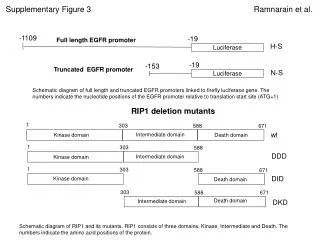

Schematic Diagrams of EGFR and RIP1 Promoters and Their Mutants Linked to Luciferase
E N D
Presentation Transcript
Full length EGFR promoter -19 -153 Truncated EGFR promoter N-S Luciferase H-S Luciferase Schematic diagram of full length and truncated EGFR promoters linked to firefly luciferase gene. The numbers indicate the nucleotide positions of the EGFR promoter relative to translation start site (ATG+1) -1109 -19 1 303 588 671 RIP1 deletion mutants wt Intermediate domain Death domain Kinase domain 1 303 588 DDD Intermediate domain Kinase domain 1 303 588 671 DID Kinase domain Death domain 303 588 671 Death domain Intermediate domain DKD Schematic diagram of RIP1 and its mutants. RIP1 consists of three domains, Kinase, Intermediate and Death. The numbers indicate the amino acid positions of the protein. Supplementary Figure 3 Ramnarain et al.