OA 11.4
OA 11.4. Chapter 18 ( pp 487-496). Injuries to the Foot & Ankle. O bjectives. Identify… The bones of the foot & ankle The ligaments of the foot & ankle The muscles of the foot & ankle The tendons of the foot & ankle The blood vessels & nerves of the foot & ankle Other structures.

OA 11.4
E N D
Presentation Transcript
Chapter 18 (pp 487-496) Injuries to the Foot & Ankle
Objectives Identify… • The bones of the foot & ankle • The ligaments of the foot & ankle • The muscles of the foot & ankle • The tendons of the foot & ankle • The blood vessels & nerves of the foot & ankle • Other structures
The bones • The foot contains 28 bones • Phalanges (16) • Proximal (1-5) • Intermediate (2-5) • Distal (1-5) • Sesamoids (1)
The bones • The foot contains 28 bones • Metatarsals (5) • Tarsal bones (7) • Cuneiforms • Medial • Intermediate • Lateral • Cuboid • Navicular • Talus • Calcaneous • Tibia • Fibula
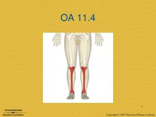
The bones • Tibia • Fibula
Phalanges • There are 14 total phalanges • Great toe = 2 • All other toes = 3 • Each toe has a proximal, intermediate, & distal phalange (except Great toe)
Phalanges • Great toe has 2 sesamoid bones • Act like “mini patella” to increase leverage and protect the joint
metatarsals • Each toe has a corresponding metatarsal (MT) • Important areas: • Head of the 1st MT • Base of the 5th MT • In-between the 2-3 MT heads
Tarsal bones • The tarsal bones make up the mid- & rear-foot • Cuneiforms (E, F, G) • Medial (G) • Intermediate (F) • Lateral (E) • Cuboid (C) • Navicular (D) • Talus (B) • Calcaneous (A)
Bones of the leg • Tibia and fibula extend past the talus bone • Distal end is referred to as the malleolus • Medial = tibial • Lateral = fibular
Bones of the leg • Tibia • Larger of the two bones • Primary weight bearing bone
Bones of the leg • Fibula • Smaller bone, extends more distally • Provides for muscle attachments • ≤ 10% weight bearing
the ligaments • Important ligaments in the foot: • Calcaneonavicular ligament (spring ligament)
The ligaments • Medial ankle: • Deltoid ligament
The ligaments • Lateral ankle: • Anterior talofibular (ATF) • Calcaneofibular(CF) • Posterior talofibular(PTF) • Not shown
The ligaments • Lower leg: • Distal anterior tibiofibular ligament • Distal posterior tibiofibular ligament • Interosseous membrane
OA 11.5 • Identify the bones: • A • D • E • C
articulations • Interphalangeal (IP, PIP, DIP) • Metatarsophalangeal (MP or MTP) joints • Intermetatarsal joints • Tarsometatarsal (TMT) joints • Subtalar joint • Talocrural joint
Subtalar joint • Articulation of the talus & calcaneus
Talocrural joint • Tibia, fibula, talus • Mortise & tenon configuration • Talus is wedge-shaped • Lateral malleolus extends more distally than medial malleolus
Arches of the foot • Support body weight • Absorb forces from the ground • Provide space for blood vessels, tendons, & muscles
Arches of the foot • Metatarsal Arch – across metatarsal heads • Transverse Arch – across metatarsal bases & cuneiforms • Medial Longitudinal Arch – along the medial aspect • Lateral Longitudinal Arch – along the lateral aspect
Plantar fascia • Broad, thick tissue covering the bottom of the foot • Extends from the calcaneus to the base of each metatarsal • Supports the foot against downward forces
Muscles & tendons • Intrinsic muscles of the foot: • Toe extensor • Toe flexors (3) • Great toe & 5th toe abductors • Great toe adductor
Muscles & tendons • Extrinsic muscles of the foot: • Divided by compartments • Anterior • Lateral • Superficial posterior • Deep posterior
Muscles & tendons • Anterior compartment • Tibialis anterior • Extensor hallucislongus • Extensor digitorumlongus • Dorsiflex the foot
Muscles & tendons • Lateral compartment • Peroneus longus • Peroneus brevis • Evert the foot
Muscles & Tendons • Superficial posterior compartment • Gastrocnemius • Soleus • Plantaris • Plantarflex the foot
Muscles & tendons • Deep posterior compartment • Tibialis posterior • Flexor hallucislongus • Flexor digitorumlongus • Plantarflex & invert the foot
Neurological & Vascular • Tibial nerve • Posterior leg & plantar aspect of foot • Common peroneal nerve • Anterior leg & foot • Blood supply • Anterior tibial artery Dorsal pedal artery • Posterior tibial artery
OA 11.12 If an athlete came to you complaining of ankle pain, how would you address them? • What questions would you ask to gather clues about what is going on? • What are some relevant observations to make regarding their body?
The Foot & Ankle Evaluation
Objectives Identify… • Pertinent information to gather during a foot & ankle evaluation • Important observations to make during a foot & ankle evaluation ???
The Secondary Survey • After ruling out life-threatening injuries, we begin the secondary survey • Treat for major injuries with acute on-field care • Begins with an assessment of vital signs • Musculoskeletal Assessment DOCUMENT EVERYTHING!
The Evaluation Process H.O.P.S. H.I.P.S. History Inspection Palpation Range of Motion Special tests • History • Observation • Palpation • Range of motion • Special tests
History • What happened? • Gain information about the patient and the injury • Most critical part of the evaluation! • Past medical history • History of the present condition
History • Start with generic history questions • Chief complaint • Age • Occupation / sport / position etc. • General healthcondition • Activity level • Medications
History • History of previous injuries • What happened? • Who did you see? • What did they tell you? • How long were you out? • Has it fully resolved?
History • Mechanism of injury • How did it happen? Tension = sprain; fracture; strain Torsion = sprain; fracture Compression = contusion; fracture Shear = fracture; sprain Bending = fracture
History Ask these questions regarding PAIN • P-rovocation – what causes it? what makes it better? • Q-uality – what does it feel like? neurological symptoms? • R-egion – where does it hurt? can you point w/one finger? • S-everity – how bad does it hurt? (1-10) • T-iming – when does it hurt? how long?
History • Sounds & sensations • Did you hear any sounds? Did you hear any pops, crackles, snaps, clicks? • What could this indicate??? • Did you feelanything unusual?
History • Specific to the foot & ankle • Previous history = chronic ankle instability • Mechanism of injury = ROM (Inversion, Eversion, Plantarflexion, Dorsiflexion) • Location of pain – heel, foot, toes, arches, lateral ankle, medial ankle, etc. • Determines what is injured • Changes in activity,footwear, or training surfaces
Observation • Athlete Moving? • Position of athlete? • Conscious? • Primary Survey • Inspect injury site • Secondary Survey