Download
1 / 73
870 likes | 1.49k Vues
Management of liver diseases in pregnancy. Moderator-Prof. Anoop Saraya Candidate-Dr. Moka Praneeth. Contents. Pregnancy – physiologic changes Hepatitis-E Hepatitis-B Acute liver failure Cirrhosis & Portal hypertension ICP HG HELLP syndrome AFLP.

E N D
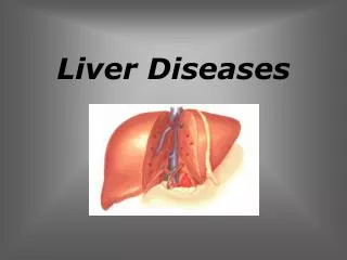
Management of liver diseases in pregnancy Moderator-Prof. AnoopSaraya Candidate-Dr. MokaPraneeth
Contents • Pregnancy – physiologic changes • Hepatitis-E • Hepatitis-B • Acute liver failure • Cirrhosis & Portal hypertension • ICP • HG • HELLP syndrome • AFLP
Physiological changes in liver tests during normal pregnancy
Liver diseases in pregnancy coincidental with pregnancy Only in the setting of pregnancy Chronic liver diseases e.g.: cholestatic liver disease, autoimmune hepatitis, Wilson disease, viral hepatitis, etc… not associated with preeclampsia Preeclampsia- associated The preeclampsia itself • Hyperemesis • gravidarum HELLP-syndrome • Intrahepatic cholestasis • of pregnancy AFLP
Hepatitis E virus infection and fulminant hepatic failure during pregnancy • 50 pregnant and 50 non-pregnant women with FHF and 150 pregnant healthy females without liver disease as controls were recruited for the study. • Serologically (38/50; 76%) as well as by RT-PCR (28/50; 56%), a significantly higher HEV positivity rate was found in pregnant FHF patients compared to non-pregnant women (serologically 15/50; 30%; RT-PCR 7/50; 14%). Jilani N et al. J GastroenterolHepatol. 2007
Hepatitis E virus infection and fulminant hepatic failure during pregnancy • CD4 counts were lower (P < 0.05), while CD8 counts were higher (P < 0.05), and their ratio (CD4/CD8) in HEV positive pregnant FHF patients was significantly lower (P < 0.01) when compared to that of HEV negative pregnant FHF women or controls. • Levels of estrogen, progesterone and beta-HCG were also found to be higher (P < 0.001) in HEV positive pregnant FHF patients when compared to HEV negative patients or controls. • HEV infected pregnant FHF patients had a significantly higher mortality rate of 65.8% (25/38) compared to 23.5% (4/15) in HEV positive non-pregnant women (P < 0.001
Immunological alterations in pregnant women with acute hepatitis E. • Pregnant women with HEV had generalized immune suppression characterized by decrease in lymphocyte response to phytohemagglutinin(PHA) with a predominant Th2 bias as compared to non pregnant women with hepatitis E and normal healthy controls. • Neither normal healthy pregnant women nor nonpregnant HEV infected women demonstrated decreased response to PHA. Pal R et al. J GastroenterolHepatol. 2005
Termination of pregnancy in HEV-ALF? Banait VS et al. Indian J Gastroenterol 2007
Treatment algorithm for an HBV-infected woman who is already on antiviral therapy and presents with an unexpected pregnancy
Impact of pregnancy on chronic HBV • No worsening of liver disease in majority • Overall increase in HBV DNA levels during pregnancy • Median ALT levels decreased during pregnancy • Increase in ALT (3 x lowest ALT) within 6 months after delivery • Case reports of postpartum hepatic exacerbations Terrault et al. Semin Liver Dis. 2007 Soderstrom et al. Scand J Infect Dis 2003 Borg et al. J viral Hep 2008 Rasheed et al. Int J GynaecolObstet 2013
HBV Infection in Women Considering Starting a Family: Which Drug? • FDA classification: based on in vitro and animal studies • Pregnancy class B: telbivudine and tenofovir DF • Pregnancy class C: interferon, adefovir, entecavir, and lamivudine • Human data: • Antiretroviral pregnancy registry: safety established for lamivudine and tenofovir, including exposure in first trimester[1] • Clinical studies of antiviral therapy to prevent perinatal transmission: safety established for lamivudine and telbivudine, mainly exposure in third trimester[2-5] 1. Antiretroviral Pregnancy Registry. December 2012. 2. Xu WM, et al. J Viral Hepat. 2009;16:94-103. 3. Shi Z, et al. Obstet Gynecol. 2010;116:147-159. 4. Han GR, et al. J Hepatology. 2011;55:1215-1221. 5. Pan CQ, et al. Clin Gastroenterol Hepatol. 2012;10:520-526.
Incidence of Birth Defects With in Utero Exposure to HBV Nucleos(t)ide Analogues • Data derived from Antiretroviral Pregnancy Registry, 1/1989 - 7/2012[6] • International, voluntary, prospective, exposure-registration cohort • Data on exposure in HBV-monoinfected mothers began in 1/2003 • Metropolitan Atlanta Congenital Defects Program, a population-based birth defects surveillance program administered by CDC[6,7] • Overall birth defects: 2.72% (95% CI: 2.68-2.76) 6. Antiretroviral Pregnancy Registry. December 2012. 7. Correa A, et al. Birth Defects Res A Clin Mol Teratol. 2007;79:65-186.
Prevention of Perinatal HBV Transmission • Cornerstone: HBIG + HBV vaccine • HBIG + first dose vaccine within 12 hrs of birth, different sites • Efficacy: ~ 95% • Reasons for failure • Delay in administration of HBIG and first dose of vaccine • Failure to complete vaccine series/Poor quality vaccine • Mother HBeAg positive and/or high HBV DNA • In utero infection • HBsAg mutation/Escape mutants • Immunocompromised host 1. Lok AS, et al. Hepatology. 2009;50:661-662. 2. Mast EE, et al. MMWR Recomm Rep. 2005;54(RR-16):1-31.
Meta-analysis of Lamivudine to Interrupt Perinatal Transmission of HBV Risk Ratio (95% CI)* 100 1 0.1 0.01 10 Favors Lamivudine Favors Control Risk Ratio (95% CI)* *Risk ratio calculated using the Mantel-Haenszel random-effects model. 1 0.1 100 0.01 10 Favors Lamivudine Favors Control 17. Han L, et al. World J Gastroenterol. 2011;17:4321-4333.
Algorithm for HBV Management in Women During Pregnancy Pregnant women with HBV infection Active disease/suspected cirrhosis: consider initiating treatment with tenofovir 1st trimester: assess HBV replication and liver disease End of 2nd trimester: quantitative HBV DNA and ALT levels HBV DNA < 106 IU/mL* HBV DNA > 106 IU/mL* Consider initiating treatment with tenofovir, lamivudine, or telbivudine at 28-32 wks† Monitor;infant receives HBIG + vaccine at birth Infant receives HBIG + vaccine at birth *The cut-off level of maternal HBV DNA level for initiation of therapy is unclear, and HBV DNA from 6-8 log10 IU/mL can be considered for therapy based on physician and patient preference. †Tenofovir is preferred if treatment is expected to be > 12 weeks or if treatment is expected to continue while breastfeeding.
Pregnant Women With High HBV DNA and Not Initially on Antiviral Therapy • When to stop antiviral after delivery? • To prevent perinatal transmission: immediately, especially if mother plans to breast-feed, or up to 3 mospostdelivery • To treat liver disease: continue until therapeutic endpoint • What is the risk of posttreatment flare? • Seemingly rare, but mild ALT elevation common; also seen in postpartum period for women not receiving antiviral • Decompensation not reported in clinical trials; likelihood low because most pregnant women have early-stage liver disease • Important to closely monitor ALT after antiviral therapy is discontinued (eg, 1, 3, and 6 mosposttreatment) 19. Ter Borg MJ, et al. J Viral Hepat. 2008;15:37-41.
Cesarean vs Vaginal delivery in CHB- a meta-analysis • Infant serum Anti-HBs postivity at birth (RR= 1.24, 95% CI 0.89-1.74, p = 0.2) or at 6-7 months (RR= 0.98, 95% CI0.86-1.11, p = 0.73) was not significantly different • The incidence of infant CHB infection may have been higher in the vaginal delivery group (R=2.2, 95% CI 1.02-4.74, P = 0.04) Xu et al. Dig Dis Sci. 2014
Acute HBV in pregnancy Transmission rates: • 10% in early pregnancy • 60% at or near time of delivery
ALF in pregnancy • 1015 consecutive patients of ALF in reproductive age group, admitted in AIIMS from January 1986 to December 2006 • 249 (38.5%) were pregnant females • The mortality rate of pregnant women and girls (53.8%) was similar to nonpregnantwomen and girls (57.2%), and men and boys (57.9%); P = 0.572. Bhatia V, Acharya SK et al. Hepatology 2008
ALF in pregnancy • A significantly higher proportion of ALF was attributable to HEV among pregnant women (59.4%), as compared with nonpregnantwomen (30.4%), and men (23.1%) (p < 0.001) • The outcome of HEV-related ALF was independent of the sex and pregnancy status of the patients (P = 0.103).
ALF in pregnancy • Mortality in HEV-ALF and non-HEV-ALF patients in pregnant women and girls was 51% (74/145) and 54.7% (52/95)(P > 0.1), respectively. • The outcome of pregnant ALF patients was also unrelated to the trimester of pregnancy. • The mortality of non-HEV-related ALF among the pregnant women and girls (54.7%), age-matched nonpregnant women and girls (61.7%), and men and boys (62.8%) were also similar (P > 0.1). Bhatia V, Acharya SK et al. Hepatology 2008
Induction of delivery in ALF with IUD • Increased risk of peripartum hemorrhage due to associated coagulopathy 1 • rFVIIa is a useful adjunct to standard management in postpartum hemorrhage secondary to acute liver failure of pregnancy-related liver disorders.1 • When a dead fetus has been in utero for 3-4 weeks, fibrinogen levels may drop, leading to a coagulopathy.2 • Does a dead (< 2 weeks) fetus lead to sepsis? • Should we routinely remove the dead fetus in pregnant women with ALF with IUD? Goel a et al. Indian J Gastroenterol. 2013 Jul Tempfer CB et al. J Womens Health (Larchmt). Apr 2009
CLD- pregnancy • Preexisting varices : • 78% GI bleeding during pregnancy • T2/T3 as maternal blood volume maximally expanded and fetus causes ↑ compression of IVC collateral vasculature • Mortality rate of 18% to 50%. • Most patients with CLD infertile due to hypothalamic- pituitary dysfunction. Russell MA et al. Semin Perinatol 1998;22:156-165. • MATERNAL RISKS: • Variceal Bleeding ( 18-32 %) • Hepatic Decompensation(24%) • Hepatic Encephalopathy • PPH ( 7-10%) • Rupture of splenicA aneurysms(2.6%) FETAL RISKS : • ↑ risk of abortions (30-40%) • ↑ risk of prematurity / IUGR (25%) • Perinatal death (18%) Hay et al. Hepatology 2008
Treatment of variceal bleeding • Endoscopic variceal band ligation • Superior to sclerotherapy – no chemicals instilled into blood stream. -Expert opinion • Octreotide (B) • Safety in pg not determined • Could cause arterolar vasospasm • Decreased placental perfusion and increased risk of placental abruption as well as • HTN, MI, peripheral ischemia • Endoscopy • Safe when done with caution
Pregnancy and cirrhosis • Review of data from 1984 – 2009 • Kings College Hospital • 62 pregnancies in 29 women • Median MELD was 7 (range 6 – 17) • Median CPS was 5 ( range 5-8) • Live birth rate was 58% • Median gestational age 36 w Westbrook RH et al ClinGastroHep 2011;9: 694-9
Pregnancy and cirrhosis • Maternal complications occurred in 10% • Ascites • Encephaolpathy • Variceal hemorrhage • Associated with MELD > 10 • MELD predicted which patients were to have liver related complications • AUC 0.8 • 83% sensitivity and 83% specificity • No one with MELD <6 had any liver related complications Westbrook RH et al ClinGastroHep 2011;9: 694-9
Cesarean vs vaginal delivery in CLD • Experts use elective cesarean section or forceps delivery under extradural analgesia to decrease the risk of variceal rupture (no RCTs are available)1, 2 • If a prophylactic cesarean section is performed, a vascular surgeon should be available as bleeding from pelvic or abdominal wall collaterals may occur3 BenjaminovFS, Heathcote J. Am J Gastroenterol2004 Heriot JA et al. Br J Anes1996 Misra S, Sanyal AJ. Clin Liver Dis 1999
Intrahepatic Cholestasis of Pregnancy (ICP) • Incidence 0.1% - 1% of pregnancies, 20% among twins, 7.5% of pregnancies (AIIMS) • Recurrence rate of 50-70% in subsequent pregnancies • Pruritus (initially in soles & palms, only at night, and later continuously progressing to trunk and face) develops in late 2nd and 3rdtrimester
ICP • Jaundice (usually mild- bilirubin < 5 mg/dL) develops 2 weeks later, plateaus and remains constant until delivery • Pruritus worsens with the onset of jaundice • Symptoms usually abate within 2 days of delivery.
ICP-lab investigations • The most specific and sensitive marker of ICP is total serum bile acid (BA) levels > 10 µmol/L • Chenodeoxycholic acid and cholic acid ↑ sed 10 times N( > 11µmol/L), ↑ cholic acid % ( > 42 %), ↓ glycine/ taurine bile acid ratio (<1) • ALT, AST- mild to 10-25 fold increase, SAP- increased upto 4 fold • GGT-mildly elevated in 30%, more likely in those with genetic component • Glutathione s- transferase α - new marker being investigated
MATERNAL RISKS FETAL RISKS • Prematurity: ↑ iatrogenic prematurity ( 7- 25 % ) vs ( 4- 12 %) . • Passage Of Meconium : • preterm than in term (25% vs 12%) • preterm controls (18% vs 3%). • mc if BA > 40 umol/l vs 20 • Risk ↑ linearly ( 19.7% ↑ for each • 10 umol/l ↑ BA conc.) . • Intrapartum Fetal Distress • Kenyon et al.. BJOG 2002; 109 • Higher Caesarean Rates (10 – 36 %) → ↑ RDS & TTN • Stillbirth: (1.5 %) to that of general • population (0.5%). • RCOG 2011 • Deposition of bile acids in the skin → pruritus → dermatitis artefacta. • Hepatic impairment → prolonged prothrombin time → post partum hemorrhage RCOG 2011 Arrese. Annals of Hepatology 2006; 5 (3): 216-218 • IUD seems to be due to – • acute anoxia (placental chorionic vein • spasm , umbilical vein spasm) • umbilical vein constriction • fetal cardiac arrest ( infiltration of fetal • cardiomyocytes by bile salts) • Kenyon et al. BJOG 2002 • Gorelik et al. BJOG 2006
T. UDCA 15 mg/kg/d (or > 1 g/day) (– drug of choice) reduces pruritus, ↓ total serum bile acids, ALT values and bilirubin levels, improves cholic/CDCA ratio • Other agents with limited benefit: Phenobarbital 100 mg OD, Hydroxyzine, SAME 1600 mg OD, Cholestyramine- takes 2 weeks to work, Aluminium containing antacids, Dexamethasone 12 mg QID for 7 days
Investigations for HG • Urinalysis for ketones & specific gravity • Serum electrolytes: Na, K, Cl • LFTs- Elevated transaminases (50-65%) upto 200 IU/L, SAP upto 2 times, serum bilirubin upto 4 mg/dL- makes it a liver disease? • Serum amylase & lipase upto 5 times • TSH, FT4, Urine culture, Hct, screening for HAV, HBV, HCV • Obstetric & abdominal USG
Management of HG • i.v. fluids (NS, RL) over 2-3 hours • Thiamine/vitamin B1 • Enteral/Parenteral nutrition • Separating solids & liquids, Eating small frequent meals of bland foods, Avoiding fatty foods (eg: Potato chips) • Avoid drinking cold/sweet beverages • Eliminate pills with iron; give high protein snacks
COMPLICATIONS Maternal complications Fetal complications No increased risk of congenital malformations . Growth restriction. Wernicke’s encephalopathy is associated with a 40% incidence of fetal death. • Hypokalemia - lethargy, skeletal muscle weakness and cardiac arrhythmias • Hyponatremia and CPM. • Vitamin B6/B12 def • Malnutrition. • Mallory-Weiss oesophageal tears. • Wernicke’s encephalopathy. • Venous thromboembolism . • Psychological morbidity. NICE 2010
HELLP Syndrome • 1 in 1000 , 10-20 % of severe preeclampsia / eclampsia • Most common in white, multiparous and older women
Clinical Presentation SYMPTOMS
Clinical Presentation signs
Laboratory Diagnostic Criteria for HELLP syndrome Haemolysis Abnormal peripheral smear : spherocytes, schistocytes, triangular cells and burr cells Total Bilirubin level > 1.2 mg/dL Lactate dehydrogenase level > 600U/L Elevated liver function test result Serum aspartate amino transferase level > 70U/L Lactate dehydrogenase level >600 U/L Low platelet count(Most reliable indicator) Platelet count < 150 000/mm3 • Requires at least 2 of above mentioned abnormalities