
The Small Intestine: Anatomy, Function, and Digestion Process
Explore the intricacies of the small intestines, where the majority of nutrients are absorbed into the bloodstream. Discover the anatomy shared by monogastrics and ruminants, including the divisions into duodenum, jejunum, and ileum. Dive into the small intestinal wall's structure and learn about the role of the nervous system in intestinal motility. Understand the digestion process in the small intestine and the breakdown of carbohydrates, proteins, and fats. Uncover how electrolytes, water, and vitamins are absorbed across the intestinal wall and the importance of enzymes in this process.

The Small Intestine: Anatomy, Function, and Digestion Process
E N D
Presentation Transcript
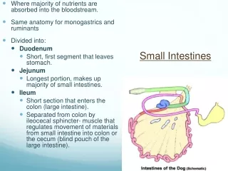
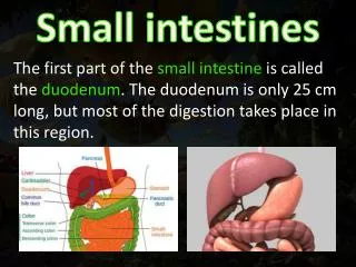
Small Intestines • Where majority of nutrients are absorbed into the bloodstream. • Same anatomy for monogastrics and ruminants • Divided into: • Duodenum • Short, first segment that leaves stomach. • Jejunum • Longest portion, makes up majority of small intestines. • Ileum • Short section that enters the colon (large intestine). • Separated from colon by ileocecal sphincter- muscle that regulates movement of materials from small intestine into colon or the cecum (blind pouch of the large intestine).
Small Intestinal Wall • Similar to other segments of GI tract. • Mucosa, submucosa,muscularis, serosa • Mucosa – increased surface area due to many folds and villi • Each villus contains thousands of microvilli (brush border) • Microvilli – have both digestive enzymes and carrier molecules for nutrients, vitamins, and minerals embedded in cell membranes • Crypts- invaginations of mucosa around each villus. New cells are produced and pushed from bottom of crypt to replace cells of villus • Goblet cells present. Mucus helps with flow of ingesta.
Nervous System and Small Intestines • Parasympathetic nervous system provides stimulation for intestinal motility, secretions, and blood flow. • Sympathetic nervous system decreases circulation to the intestines. • Intestinal tract is constantly functioning and is never at rest.
Small intestine Motility • Peristalsis • Circular contractions prevent backflow of ingesta, longitudinal muscles propel ingesta caudally • Dilation of bowel with ingesta stimulates peristalsis • CCK and Prostaglandins can both affect motility. • Fats/protein in the intestine stimulate the mucosa to release CCK, which increases intestinal motility (opposite of the effect on the stomach) • Prostaglandins can increase GI motility and secretions which can lead to colic. • Segmental contractions slow the movement of ingesta to allow time for it to be both mixed with intestinal enzymes and absorbed through the intestinal wall. • Many times diarrhea is caused not due to increased peristalsis, but lack of segmental contractions.
Small Intestine Digestion • Electrolytes, water, and vitamins • Absorbed intact across SI wall • Carbohydrates, proteins, and fats • Chemically digested via enzymes in the lumen and enzymes on the microvilli b/c they are too large to pass through the mucous membrane
Carbohydrate Digestion • Starch is broken into disaccharides by amylase found in the saliva and from the pancreas • Disaccharides are broken down into monosaccharides by enzymes (lactase, sucrase, and maltase) in microvilli • Monosaccharides can then be transported across microvilli and absorbed into blood • Microvilli enzymes are dependent on diet • Lactose-intolerant animals/diarrhea Food Enzyme Source Broken into Fate Starch Amylase Saliva, Pancreas Disaccharides lactose sucrose maltose Lactase Brush border Monosaccharides Sucrase glucose Absorbed Maltase galactose Absorbed fructose Absorbed
Protein Digestion • Protein chains (1000’s of amino acids) are broken into smaller polypeptides (100’s of amino acids) by pepsin • Polypeptides are broken down into peptides (several amino acids) by pancreatic proteases • Peptides are broken down into amino acids, dipeptides, and some tripeptides by peptidases are then absorbed Food Enzyme Source Broken into Fate Protein Pepsin Stomach Polypeptides Proteases SI (Pancreas) Peptides Peptidases Brush border Amino acids Absorbed di-peptides Absorbed tri-peptides Absorbed
Fat Digestion • Agitation of the pyloric antrum emulsifies (breaks down) fat globules (triglycerides) into smaller droplets • Bile acids from the liver coat the fat droplets in duodenum • Keeps them from re-forming into globules again • Arranges them to make them more water soluble • Pancreatic lipases (fat-digesting enzymes) penetrate bile acid coating • Digest triglycerides to form glycerol, fatty acids, and monoglycerides • Bile acid + lipid = micelle, which is absorbed through the microvilli • Vitamins A, D, E, K are often absorbed with the micelles Food Enzyme Source Broken into Fate Lipids Bile acids SI (Liver) small fat droplets Lipases SI (Pancreas) glycerol Absorbed fatty acids Absorbed monoglycerides Absorbed micelles Absorbed
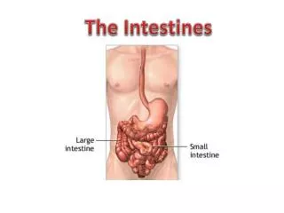
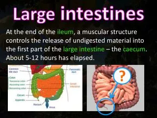
Large Intestine • Species variation in structure • Components 1. Cecum - blind sac at ileocecal junction 2. Colon 3. Rectum • Primary functions - • Store feces until they can be eliminated (carnivore) • Recover fluid and electrolytes (carnivore) • Hindgut fermentation (non ruminant herbivores) • Equine, guinea pigs, rats, rabbits, swine
Large Intestines • Carnivores: simple, tubular colon; poorly developed cecum • Nonruminant herbivores: very large colon and cecum (hindgut) • Fermentation site • Modifications of cecum and colon allow fermentative digestion in hindgut • similar to rumen • VFA’s (produced by microbes) absorbed from cecum and colon for energy needs (similar to rumen) • Possible areas of impaction • Flexures, Small colon • Cause of colic
Horse Hindgut • Consists of 4 sections: • Cecum, Ventral colon (right and left halves), Dorsal colon (right and left halves), Small colon • Cecum is composed of: • Base, Main body, Apex • Cecum and dorsal and ventral colons have longitudinal bands that separate the structure into a series of sacs called haustra • Cecum is separated from colon by cecocolic orifice • The role of the small colon is to absorb electrolytes, water, and any VFA’s that were not previously absorbed. Ileocecal sphincter►Cecum►Right ventral colon►sternal flexure►left ventral colon►Pelvic flexure►left dorsal colon►diaphragmatic flexure►Right dorsal colon►small colon
Rectum • Terminal portion of the large intestine; an extension of colon • Capable of more expansion than colon • Mucus-secreting glands lubricate feces to aid their passage • Has sensory receptors that detect stretching or distention and stimulates defecation response.
Anus • Composed of: • Internal sphincter under autonomic control • (Parasympathetic system causes relaxation, Sympathetic system causes constriction) • External sphincters under voluntary control • As rectum distends, stretch receptors cause partial relaxation of internal sphincter. Fecal material moves into the Internal Sphincter Canal which stimulates more stretch receptor increasing urge to defecate. • Stretching of Anal mucosal receptors increase the sense or need for defecation • Surgery or disease in anal region can damage sphincter muscles and nerves, causing incontinence
Liver’s Role in the GI Tract • Filters, stores, and/or metabolizes materials absorbed from GI tract before they reach blood. • Removes toxins, infectious agents, old blood cells that enter the body via the GI tract. • Glucose, amino acids, and vitamins are stored or metabolized. • Glucose absorbed by the GI tract can be stored in the liver as glycogen (glycogenesis). When glucose is needed in the blood, glycogen is broken down by the liver (glycogenolysis). Gluconeogenesis is the process of glucose being made in the liver by using amino acids. • Major source of blood proteins • Albumin
Gallbladder • The liver produces bile which contains bile acids, cholesterol, and bilirubin • Bile is secreted into bile ducts, which lead to the hepatic duct, which leads to the gallbladder for storage (not horse) • The gallbladder stores bile until it is stimulated by CCK (due to fat in SI), causing it to contract. • Contraction forces bile down the common bile duct into the duodenum, where it aids in the digestion of fat.
Pancreas’ Role in the GI Tract • Exocrine gland(secretes substances to outside of body through a duct) • Endocrine gland(secretes hormones directly into the blood without going through a duct) Exocrine functions: • Produces pancreatic amylase, proteases, lipase • Secretes bicarbonate (HCO3-)into duodenum • Neutralizes acidity of stomach contents and maintains pH in duodenum needed for proper enzyme function Endocrine functions: • Produces insulin and glucagon • Regulates blood glucose levels: Insulin moves glucose from the blood to the body’s tissues. Glucagon stimulates gluconeogenesis and glycogenolysis in the liver.
![STOMACH, INTESTINES, RECTUM [SURGICOSE]](https://cdn4.slideserve.com/8061806/medical-instruments-medical-instruments-dt.jpg)