Download
1 / 118
1.18k likes | 1.33k Vues
Explore the anatomy, ultrasound techniques, and diseases of the liver, gallbladder, and biliary tract. Learn about hepatic segments, portal veins, and common hepatic pathologies. Discover how to perform upper abdominal ultrasound scans using different patient positions and probe orientations, and interpret findings such as fatty liver, cirrhosis, and focal lesions. This comprehensive guide offers valuable insights into diagnosing hepatobiliary conditions using ultrasound imaging.

E N D
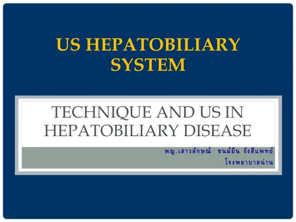
US hepatobiliary system Technique and US in hepatobiliary disease พญ.เสาวลักษณ์ ชนม์ยืน รังสีแพทย์ โรงพยาบาลน่าน
Outline • Anatomy • US technique • Hepatobiliary tract disease( Liver, gallbladder and biliary tract )
ส่วนยอด บน ของ ตับ อยู่ที่ซี่โครงที่ 5 หรือ ต่ำกว่าราวนม ครึ่งนิ้ว
ท่อที่ผ่าน เข้า-ออก Liver มี 4 ท่อดังนี้ 1.Hepatic Vein 2. Portal Vein 3. Bile Duct 4. Hepatic Artery
Upper Abdominal Ultrasonography • supine position นอนหงาย • left lateral decubitus position นอนตะแคง เอาด้านซ้ายลง • right lateral decubitus positionนอนตะแคง เอาด้านขวาลง
วางprobeเอียงไปทางด้านซ้ายวางprobeเอียงไปทางด้านซ้าย เท้า ศีรษะ STOMACH
เอียงprobeจากด้านซ้ายมาเกือบกึ่งกลางเอียงprobeจากด้านซ้ายมาเกือบกึ่งกลาง stomach Aorta
เห็นaorta and branches aorta
เห็น pancreasในการตรวจ Sagittal view in epigastrium
เลื่อน probe ผ่านกึ่งกลาง มาทางขวา
Caudate lobe IVC
เอียงprobeขึ้นไปทางศีรษะจะเห็นหัวใจเอียงprobeขึ้นไปทางศีรษะจะเห็นหัวใจ
เลื่อน probeลงมาจะเห็น pancreas
IVa II VIII VII
MHV LHV RHV
Portal vein • Periportal fibrofatty tissue produces brighter echoes around the portal veins • Normal size ไม่ควรเกิน 13 mm ถ้ามากกว่านี้ให้สงสัยว่ามีภาวะportal hypertension
Main portal vein แยกเป็น right และ left portal veins
Gallbladder GB PV
Branches of Right Portal vein V VIII VI VII
Right subcostal section in left lateral decubitus position ท่าตะแคงจะทำให้ ตับเลื่อนลงมา เราจะเห็น lesion บางอย่างที่ ท่านอนหงาย มองไม่เห็น
Liver Size - Right MCL วัดcraniocaudal direction ขนาดน้อยกว่า 15 cm • Indirect signs of hepatomegaly extension of the right lobe below the lower pole of the kidney rounding of the inferior tip of the liver extension of the left lobe into the LUQ above the spleen
Liver abnormality Parenchymal disease -Fatty liver -Liver cirrhosis -Periductal fibrosis Liver mass /Focal lesion
Liver parenchyma • Homogeneous echogenicityโดยที่ echogenicity of the liver ≥ renal cortex, < renal capsule < spleen, < pancreas • Smooth surface • Visible tubular structuresare hepatic veins and portal veins • Hepatic veins ; thin wall IVC • Portal vein ; thick wall hepatic hilum • Bile duct parallel with portal vein seen at porta hepatis
Diffuse hepatic inhomogeneity • COMMON -cirrhosis -metastasis -fatty infiltration • UNCOMMON -hepatocellular cancer -hepatic fibrosis -lymphoma
Fatty infiltration • Intracellular deposition of triglycerides within hepatocytes • Causes – Alcoholic ,Steroid ,DM,Drug • NASH ( Nonalcoholic Steatohepatitis ) = Severe fatty liver with hepatomegaly with inflammation and fibrosis • Findings - Increased echogenicity of the liver, finer echotexture than normal liver • เทียบกับ kidney and pancreas
Fatty Infiltration • Sensitivity 60-94 % Specificity 66-95 % • Diffuse - Grading -mild = mildly increasing echogenicity -moderate = blurring of veins margins -severe = significant of posterior shadowing • Focal fatty infiltration