Download
1 / 1
10 likes | 114 Vues
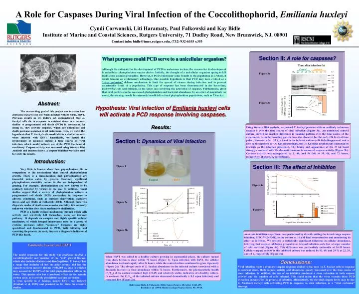
Time after infection (h). Control Infected. To 24 35 48 72 24 35 48 72. Kd. EhV1 abundance (viruses ml -1 ). 250. Infected with EhV1. E. huxleyi abundance (cells ml -1 ). 150. 100. 75. 50. 37. 0. (1). 2. (5). 4. 6. (40). Control. + EhV1. (29).

E N D
Time after infection (h) ControlInfected To 24 35 48 7224 35 48 72 Kd EhV1 abundance (viruses ml-1) 250 Infected with EhV1 E. huxleyi abundance (cells ml-1) 150 100 75 50 37 0 (1) 2 (5) 4 6 (40) Control + EhV1 (29) Photosynthetic efficiency (Fv/Fm) 8 Caspase 8 activity (RFU µg protein-1 d-1) Fold difference from control 10 12 (69) (91) 14 (54) 16 W. Wilson 1 µm Abstract: The overarching goal of this project was to assess how Emiliania huxleyi cells die when infected with its virus, EhV1. Previous results in Dr. Bidle’s lab demonstrated that E. huxleyi cells die in response to nutrient stress in a manner similar to programmed cell death (PCD) in metazoans. In doing so, they activate caspases, which are ubiquitous cell death proteases common in all metazoans. Here, we tested the hypothesis that E. huxleyi cells would die in a similar manner when infected with EhV1. Specifically, we tested the involvement of caspases during a time course of viral infection, which would indicate use of the PCD biochemical machinery. Caspase activity was measured using Western Blot Analysis and enzyme assays. A caspase inhibitor was also used to verify the results. Introduction: Very little is known about how phytoplankton die in comparison to the mechanisms that control phytoplankton growth. There is a misconception that phytoplankton are immortal unless eaten by grazers. However, significant phytoplankton mortality occurs in the sea independent of grazing. For example, phytoplankton are now known to be routinely infected by viruses in the sea. In addition, recent studies suggest that a variety of phytoplankton activate a programmed cell death (PCD) mechanism in response to adverse conditions, such as nutrient deprivation, oxidative stress, and age (Bidle & Falkowski 2004). Although these two disparate modes of death cause massive mortality, it is currently unknown whether they share mechanistic similarities. PCD is a highly refined mechanism through which cells actively and selectively kill themselves, using an intrinsic pathway. It depends on complex and highly specific cellular machinery, of which integral importance rests on a group of cysteine proteases called “caspases.” Caspases are highly specialized and fundamental to PCD, both initiating and executing the process. As such, they are a diagnostic indicator of PCD-like death. Emiliania huxleyi and EhV1 The model organism for this study was Emiliania huxleyi, a coccolithophorid and member of the “red” plastid lineage, which also includes diatoms and dinoflagellates. E. huxleyi has a range that includes all but the polar oceans, and has the capacity to form massive blooms (>100,000 sq. km.) in which it may account for 80-90% of the total phytoplankton cells in the water. This species also has a profound effect on the oceanic carbon cycle, as it actively precipitates calcium carbonate. A virus specific to E huxleyi, named EhV1, was isolated (Bratbak et al. 1993) and provided to Dr. Bidle for research studies. A Role for Caspases During Viral Infection of the Coccolithophorid, Emiliania huxleyi Cyndi Corwonski, Liti Haramaty, Paul Falkowski and Kay Bidle Institute of Marine and Coastal Sciences, Rutgers University, 71 Dudley Road, New Brunswick, NJ. 08901 Contact info: bidle@imcs.rutgers.edu, (732) 932-6555 x393 Section II:A role for caspases? What purpose could PCD serve to a unicellular organism? Although the rationale for the development of PCD in metazoans is clear, the reasons for its development in unicellular phytoplankton remain elusive. Initially, the thought of a unicellular organism opting to kill itself seems counter-productive. However, if PCD could incur some benefit to the population as a whole, it would become an evolutionary advantage. One possible hypothesis is that PCD may have evolved as a “virus exclusion” defense mechanism to limit the spread of viruses during infection and to prevent catastrophic death of a population. This type of response has been demonstrated in the bacterium, Escherichia coli, and humans, in the latter case involving the activation of caspases. Furthermore, given that viral particles in the sea exceed phytoplankton and bacterial abundance by an order of magnitude (or more), this strategy would be extremely beneficial to clonal phytoplankton populations, such as E huxleyi. Figure 3a Figure 3b Hypothesis: Viral infection of Emiliania huxleyi cells will activate a PCD response involving caspases. Results: Using Western Blot analysis, we probed E. huxleyi proteins with an antibody to human caspase 8 over the time course of viral infection (Figure 3a). An uninfected control culture showed no marked difference in banding pattern over the time course of the experiment. A similar banding pattern was also observed for the early (24 h) viral time point. However, after 35 h, a band in the infected culture (~50 Kd) disappeared, and a new band appeared at ~37 Kd. Interestingly, this 37 Kd band dramatically increased in intensity as the infection proceeded. The timing and appearance of the 37 kd band strongly correlated with the dramatic increase in measured caspase activity (Figure 3b). Caspase activity was upregulated by 5, 40, and 54 fold at 35, 48, and 72 hours, respectively, (Figure 3b, parentheses). Section I:Dynamics of Viral Infection Section III:The effect of inhibition Figure 1 Figure 4a Figure 2a Figure 4b Figure 2b An in situ inhibition experiment was performed by directly adding the broad range caspase inhibitor, FITC-VAD-FMK, to the culture at 10 µM final concentration and monitoring its effect on infection. We detected a statistically significant difference in cellular abundance, inferring that caspase inhibition prevented or delayed infection such that a larger number of cells survived (Figure 4a). This difference was particularly noticeable at 24-35 hours. Cellular caspase activity in the inhibited culture was reduced by 91, 69, and 29 % at 22, 35, and 48 h, respectively (Figure 4b). When EhV1 was added to a healthy culture growing in exponential phase, the culture turned from dark brown to clear within 72 hours (Figure 1). Upon infection with EhV1, the cellular abundance declined rapidly after 24 hours, while the control culture continued to grow rigorously (Figure 2a). The abrupt crash of E. huxleyi abundance in the infected culture correlated with a dramatic increase in viral abundance within 72 hours. Furthermore, the photosynthetic health (Fv/Fm) of the control remained high (>0.45) and relatively stable, indicative of a healthy culture. In contrast, the Fv/Fm of the infected culture decreased dramatically (<0.3 upon infection) and remained low (Figure 2b). Conclusions: Viral infection elicits a dramatic caspase response similar to that seen in E. huxleyi cells in response to nutrient stress.Both caspase activity and abundance greatly increased over the time course of viral infection. In addition, the use of an inhibitor produced a clear reduction in both caspase activity and the number of cells infected. This could mean that the virus recruits these PCD execution enzymes for the process of infection. Alternatively, the elevated caspase levels may be due to Emiliania huxleyi cells activating PCD in response to viral infection, as a “viral exclusion” strategy. References: Bidle & Falkowski (2004) Nature Reviews Microbiol. 2:643-655. Bratbak et al. (1993) Marine Ecology Progress Series. 93: 39-48.