Download
1 / 48
490 likes | 625 Vues
Learn about the characteristics, innervation, twitch duration, and regulation of skeletal muscle fibers. Understand the roles of ATP, myosin, actin, and calcium in muscle contraction and metabolism. Discover how different myosins impact muscle performance and fatigue.

E N D
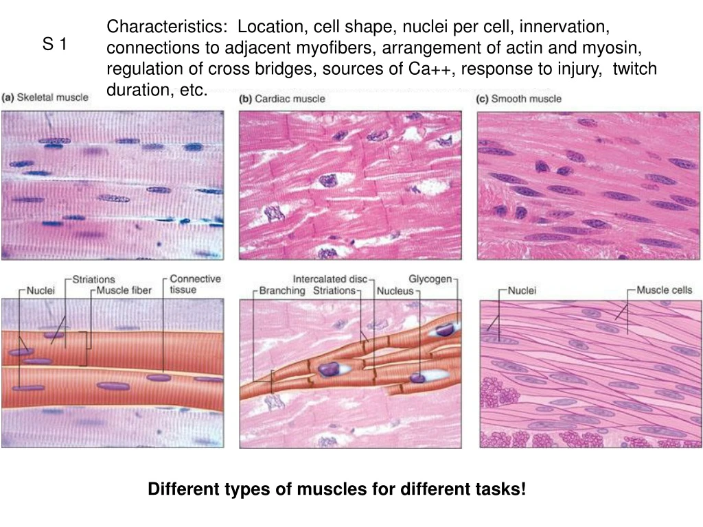
Characteristics: Location, cell shape, nuclei per cell, innervation, connections to adjacent myofibers, arrangement of actin and myosin, regulation of cross bridges, sources of Ca++, response to injury, twitch duration, etc. S 1 Different types of muscles for different tasks!
S 2 Synonyms: NMJ = neuromuscular junction Myoneural junction Motor end plate
Fig. 09.15 S 3 1 AP in motor axon releases sufficient ACh for 1 AP in skeletal muscle. Nicotinic EPP Myasthenia gravis and loss of nAChRs
Terms: Myofiber, myofibril, myofilament S 4 Fig. 09.11b Thick myofilament = myosinThin myofilament = actin
Fig. 09.12 S 5 High fAP leads to accumulation of Ca++ in sarcoplasm because Ca++ ATPase doesn’t return all Ca++ to SR quickly enough. The concentration of free calcium is directly related to force of contraction in skeletal muscle Thus we need to understand the cellular mechanism of contraction… cell biology flashbacks…
YouTube VideosRole of ATP in muscleCreepy Muscle demoSarcomere contractionPowerstoke in muscle I S 6
Fig. 09.08 S 7 Two roles of ATP: 1) Energy for powerstroke & 2) Necessary for detaching myosin from actin Crossbridge cycling continues as long as ATP and Ca++ present.
Classes of Myofibers based on Twitch Duration S 8 Each skeletal muscle fiber express only one of two different myosins isozymes: • Fast twitch = rapid hydrolysis of ATP means crossbridges cycle faster • Slow twitch = slower hydrolysis, isozyme catalyzes the reaction slower Isozymes not modified by athletic training! Contraction velocity also affected by load!
Fig. 09.02 S 9 dArk band = aligned myosin filaments lIght band = absence of myosin filaments Myofilaments
Fig. 09.03 S 11
Fig. 09.04 S 12
Fig. 09.05a S 13
Fig. 09.05b S 14 Contracted
Fig. 09.05a S 15 Image if the sarcomeres were stretched? How would the number of cross bridges capable of binding to actin be affected? How would the overlap affect the tension produced?
1QQ # 13 Answer one. • Suppose there was a mutation that rendered troponin incapable of binding Ca++. Would that embryo survive? Why or why not? • What would happen if you injected Ca++ into a skeletal myofiber? • For our twitch recordings in lab, what events (in the proper sequence) were occurring during the latent period? • Why is peak tetanic tension 3-5 times greater than peak twitch tension? How is Ca++ involved?
Fig. 09.16 S 16
Length-tension Relationship S 17 So….. Tension produced by a single myofiber varies depending on sarcomere length.
S 18 Muscle kinetics Link to cytosolic calcium concentration, release, and reuptake?
Fig. 09.20 S 19 Why does this plateau? So….. Tension produced by a single myofiber varies depending on frequency of Action Potentials. Why is peak TETANIC tension so much greater than peak TWITCH tension?
Muscle Metabolism S 20 • Classification of Myofiber types • Speed of myosin ATPase • Metabolic sources of ATP • Fatigability
Classes of Myofibers based on Twitch Duration S 21 Each muscle fiber express only one of two different myosins isozymes: • Fast twitch = rapid hydrolysis of ATP means crossbridges cycle faster • Slow twitch = slower hydrolysis, isozyme catalyzes the reaction slower Myosin isozymes not modified by athletic training!
Classes of Myofibers based on Metabolic and Enzyme profiles S 22 • Oxidative: at peak activity rely on full aerobic cellular respiration • many mitochondria, enzymes for oxidative phosphorylation, numerous capillaries, lots of myoglobin (red) • Glycolytic: at peak activity rely on glycolysis • few mitochondria, many glycolytic enzymes, large store of glycogen, fewer capillaries, little myoglobin (white) Metabolic and Enzyme profiles CAN BE modified by athletic training!
3 Sources of ATP in muscle S 23 Powerstroking & Disconnecting crossbridges Creatine phosphate, then oxidative phosphorylation (OP) from glycogen, then OP from blood glucose, then blood fatty acids. If intense, switch to glycolysis… then take a breather… oxygen debt
Fig. 09.03 S 25 Type I Type II A Type II B
Type I Type IIA Type IIB S 27 What are the causes of fatigue? Depends on the type of activity…
S 28 Causes of fatigue • High intensity, short duration exercise • Conduction failure in t-tubules • Lactic acid accumulation • Accumulation of ADP and inorganic phosphate • Low intensity, long duration exercise • As above, and • Depletion of muscle glycogen • Low plasma glucose (hypoglycemia) • Dehydration • Control pathways: “willpower” • Common in couch potatoes
S 29 So what are the ways a muscle (consisting of many myofibers) increases tension?
Fig. 09.13 S 30 Motor unit = a single somatic motor neuron and all the muscle fibers in innervates
S 31 But each motor unit has myofibers of the same type: I or IIA or IIB.
Fig. 09.26 S 33 Relationship between recruitment and motor unit type The Size Principle Size of somatic motoneuron cell body
Increasing tension in a whole muscle S 32 • Frequency of stimulation of motor neuron • Activate larger motor units • Recruitment: activate more motor units • These factors also influence actual tension • Fiber length (length-tension) relationship • Fiber diameter • Level of fatigue (state of activity)
Types of Contractions S 34 Isotonic = Same tension Isometric = Same length Aka Lengthening contraction
Response to training S 35 • Resistance training Type II change enzyme profiles: II A to II B Type II add more actin and myosin Type II increase cross-sectional area (hypertrophy) • Endurance training • Type I increases vascularity • Type I increase number of mitochondria
Fig. 09.24b S 36 Read section of King et al., 1999 that deals with analysis of muscle biopsy material in subjects taking Andro or placebo while resistance training. What changes were expected? What changes were observed?
S 37 Consider blood flow to skeletal muscles during isometric contractions. Consider blood pressure during isometric contractions.
S 38 The benefits of using trekking poles?
S 39 Chapter 9 B Properties of Smooth Muscle How does smooth muscle differ from skeletal muscle? (innervation, membrane potentials, excitation-contraction coupling, twitch duration, fatigue, etc. (Table 9-6 p.287) What are the features of membrane potential of smooth muscle? (pacemakers and slow waves) What are the differences between single-unit and multi-unit smooth muscle?(location, spread of excitation) Who cares about smooth muscles?
Excitation-contraction coupling in Smooth Muscles Figure 9.34 S 40 from SR and influx during Action Potential or graded potential Ca++ Graded potentials result in graded contractions Slow twitch of SM due to slow action of myosin ATPase. Lack troponin Special situation: Dephosphorylation & latch bridge
Comparison of Twitch Duration S 41 Latchbridge =latch state Thankful for latch state! Crucial for long-term tension of sphincters.
Comparison of Single-Unit and Multi-Unit Smooth Muscles S 42 Slow waves and pacemaker potentials Intestinal tract, uterus, small diameter blood vessels Large airways of lungs, large arteries, ciliary muscle Often with pacemaker cells Control of membrane potential by neurotransmitters, hormones, local factors for some smooth muscles (02, NO, pH, stretch, vasodilators ….)
S 43 Cardiac Myofibers Intercalated Discs: mechanical attachments of cardiac myofibers to each other, with gap junctions (electrical synapses) to conduct AP Analogy: Falling dominoes
S 44 Plateau phase
S 45 Why no tetanic contractions of cardiac muscle?
S 46 Figure 12.17 Excitation-Contraction Coupling Calcium-induced calcium release What ends the twitch? • Ca++ channels blockers: • How and where do they work? • When are they used?
S 47 Fig. 09.06 Know this table p. 287