
Uploaded by
decker
1 SLIDES
90 VUES
10LIKES
Organization of Endoplasmic Reticulum in Developing Tissue Explained
DESCRIPTION
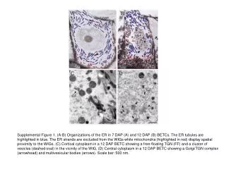
This supplemental figure depicts the ER in 7 to 12 DAP BETCs, highlighting tubules and proximity to mitochondria. It also shows cortical and central cytoplasm structures. Scale bar: 500 nm.
Download
1 / 1
Télécharger la présentation 
Organization of Endoplasmic Reticulum in Developing Tissue Explained
An Image/Link below is provided (as is) to download presentation
Download Policy: Content on the Website is provided to you AS IS for your information and personal use and may not be sold / licensed / shared on other websites without getting consent from its author.
Content is provided to you AS IS for your information and personal use only.
Download presentation by click this link.
While downloading, if for some reason you are not able to download a presentation, the publisher may have deleted the file from their server.
During download, if you can't get a presentation, the file might be deleted by the publisher.
E N D
Presentation Transcript
Supplemental Figure 1. (A-B) Organizations of the ER in 7 DAP (A) and 12 DAP (B) BETCs. The ER tubules are highlighted in blue. The ER strands are excluded from the WIGs while mitochondria (highlighted in red) display spatial proximity to the WIGs. (C) Cortical cytoplasm in a 12 DAP BETC showing a free-floating TGN (FF) and a cluster of vesicles (dashed oval) in the vicinity of the WIG. (D) Central cytoplasm in a 12 DAP BETC showing a Golgi/TGN complex (arrowhead) and multivesicular bodies (arrows). Scale bar: 500 nm.
More Related
Audio
Live Player