Comprehensive Case Taking Guidelines for Medical Professionals
This guide provides detailed instructions on conducting thorough case taking for patients, including patient particulars, presenting complaints, medical history, physical examinations, and more. It outlines the essential aspects to consider for diagnosis and management, ensuring a comprehensive approach to patient care.

Comprehensive Case Taking Guidelines for Medical Professionals
E N D
Presentation Transcript
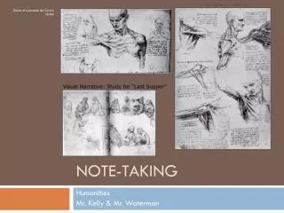
Case taking PREPARED BY , Dr. PANCHAJANI.R
Case taking • No hurry during conversation. • Hear sympathetically. • Behave patiently with the patient. • Should show good bedside manners.
Case taking includes • Particulars of the Patient • Presenting complaints • H/O Presenting complaints. • Previous History • Family history. • Personal history. • Physical examinations. • Differential diagnosis • Investigations. • Final diagnosis. • Management. • Follow up.
Particulars of the patient • Name • Age • Sex • Religion • Residence • Occupation • Social status
Presenting complaints • Main complaints with duration.( location & sensation) • Recorded in the chronological order of occurrence. • Aggravating factors & Ameliorating factors. • Accompanying symptoms.
H/O Presenting complaints • Onset of the present disease until date. • Mode of Onset of symptom.(Sudden / gradual) • Progress of the disease.( increasing/ decreasing etc..) • Related symptoms suggestive of complications of the disease. • Associated symptoms.
Previous History • Earlier diseases should be detailed in order with treatment taken. • History of earlier surgery/ trauma. • History of allergy.(medicine, food etc.) • Long term drug intake.( anti hypertensive, diabetic etc..)
Personal History • Personal habits.(smoking/drinking/addictions with duration, quantity). • Diet • Appetite. • Thirst • Weight. • Bowel& Micturition habits. • Sleep . • Females menstrual history.
Family History • Genetic History. • Diseases runs in families. • Familial diseases. (paternal/ maternal relatives) • Marital status.
Physical Examination • GENERAL PHYSICAL EXAMINATION. • SYSTEMIC EXAMINATION.
General physical examinations • Examination of a patient with out referring to a particular system. • For Proper Diagnosis and differential diagnosis.
General Physical Examination • Gait • Mental status • Built & Nutritional status • Attitude &Decubitus • Stature & posture • Facies • Pigmentations • Anaemia • Jaundice • Cyanosis • Clubbing • Oedema • Visible veins • Lymphadenopathy
Vital Signs • Pulse. • Temperature . • Respiration. • Blood pressure. • Height & weight.
Gait • Way of walking. • Important in neurological/ musculoskeletal disorder. • Note the way he moves.
Gait- Types • High stepping gait – foot is lifted high & then falls on the ground. Seen in peripheral neuritis & tabesdorsalis. • Waddling gait – abdomen is thrown forward due to lordosis.seen in pseudo hypertrophic paralysis , congenital dislocation of hip. • Spastic gait – walks the toes do not bend at knee, foot is raised by tilting up the pelvis & the leg swang forward. Stiff leg due to spasticity. Found in UMNL. • Festinant gait/ parkinsoniangaint– limbs are rigid & in a state of flexion, walks with short steps. Found in striated lesion. • Stamping gait - raises the foot high & suddently brings this down, heels first. Found in tabesdorsalis. • Reeling gait- stands on a broad base with leg wide apart, the foot is then raised high & when he walks. Reels like a drunkard
Mental status • Assess mental status & level of consciousness. • In Head injury, hepatic encephalopathy, septic shock etc.. • Grades – I - properly oriented in time, space, person. II - conscious but without orientation of time, space, person. III - Drowsy & semiconscious. IV-Unconscious but responding to painful stimuli. V- Unconscious & comatose and not responding to painful stimuli.
Built Built – structural development of body related to age & sex, race. • Under built – development poor(>food, vitamins) • Medium- average • Over build- more than normal.(hyper pituitarism, acromegaly)
Nutritional status • Nutrition- state of nourishment of the body • Proportion of soft tissue structures(muscles, soft tissues, fat) in relation to the bony structure. • Assessment – Body wt. in respect of ht.age,sex BMI(wt in kg/ ht. in m2) Triceps skin fold thickness Mid arm muscle circumferance waist hip ratio(obesity) WHR- normal- < 0.9 cm in M, < 0.85 cm in F Serum – albumin, prealbumin, transferrin, retinol binding protein.
Nutrition • Quantitative& qualitative disorder. • Quantitative- Under nutrition/ starvation- wt. loss 10% less than normal. Over wt.- gains more than 10% Obesity – more than 20% of standard wt.(BMI>30, WHR>0.9/0.85cm) • Qualitative – Malnutrition, overnutrition (food, vit.)
Body weight • Controlled by rate of energy expenditure.
Attitude • Attitude - changed position of the body or a part of the body.(posture of body as a result of disease)
Decubitus • Position of the patient in bed. • Types • Decubitus at ease • Restless D • Dorsal • Ventral • Lateral- Rt. &Lt. • Propped up • Opisthotonus • Emprosthotonus • Pleurosthotonus • Listless attitude
Stature & Posture • Stature – total height from vertex to soles. Short /Tall • Posture – Positional relationship of different regions of body. noted in standing, sitting , recumbent position. lordosis/ kyphosis /scoliosis
Facies Facial appearance & expression of pt. • Thyrotoxicosis face • Hippocratic face • Risussardonicus • Mask face • Moon face(cushingoid) • Myxoedematousfacies • Adenoid facies • Parkinsonian • Acromegaly • Mangolism
Pigmentations • Colour of skin(N-race, heriditory, climate) • Generalised/ localised • Increased/Depigmentation • Color, texture, surface of skin • Sites- face, palmar creases, skin,inside the oral cavity
Pigmentation • Increased – physiological- race, sunburn, preg. pathological- D.M, Ca, liver dis, v.vein, arsenic poisoning etc.. • Diminished – Vitiligo- patchy depigmentation Albinism – complete absence of pigmentation(skin, hair, iris , nystagmus ,impaired vision ) Leprosy- anesthetic patches
Anaemia • Quantitative/ Qualitative reduction of RBC, Hb or both below the lower limit of normal range in expected for pts. age & sex. • Manifested as pallor of mucous membrane • Sites – lower palpable conjunctiva, dorsum of tongue, inner lips, buccal cavity, nail bed, palms& soles in infants. • Normal values- • RBC- M-4.5- 6. 5 million/cubmm, F- 4-5.5mil/cu • Hb- M-14-16gm/dl, F- 13-14 gm/dl, At birth- 25gm%, • After 3 months- 20 gm%, 1 yr.- 17 gm% • MCV- 80- 94 cubic micron • MCHC- 35% • PCV – M- 40-45%, F- 38-42%
Anaemia - classification Classification- RBC volume(mcv)/Hb content. • RBC- normocytic, microcytic, macrocytic • Hb- normochromic, hypochromic • Aetiological – hypo/aplastic, dyshaemopoitic, haemolytic, post haemorrhagic • Morphological- normochromicnormocytic, N Ma cro, N micro, hypochromicmacro,hypo micro
Jaundice • Yellowish discolouration of skin, sclera, mucous membrane, & other tissues resulting from an excess of bilirubin in blood. • Normal- 0.2- 0.8mg/dl (<1mg/dl) • Conj- 0.1-0.3, Unconj-0 .2-0.7 mg/dl • Clinical jaundice > 3mg/dl • Latent jaundice -1-3mg/dl • Sites – sclera, under the tongue, soft palate, skin, palms& soles
Jaundice Types • Pre hepatic/ haemolytic • Hepato cellular/ infective • Post hepatic/ obstructive • Normal urinary urobilinogen-100-200 mg/day • Normal foecalstercobilinogen -300mg/day
CYANOSIS • Bluish/purple discolouration of skin & mucous membrane resulting from an abnormally increased amount of reduced Hb or presence of abnormal Hb in blood. • R .Hb- Exceeds 5gm/dl • Types – Central, Peripheral, Enterogenous
Cyanosis Central- CHD, emphysema, COPD etc.. • Inadequate oxygenation of blood ( cardiac/ pulmonary) • oxygen unsaturation of arterial blood(arterial hypoxaemia). • detected for tongue, inner lips, palate • extremities not affected, not cold • associated with clubbing • oxygen inhalation may relieve
Cyanosis Peripheral – excessive reduction of oxy Hb in capillaries when the blood flow is slowed down. • happens on exposure to cold ( eg. Raynaud’s phenomena) • Peripheral vasoconstriction • pts. with reduced cardiac output • Looked for nail bed, tip of nose, skin of palms &soles, ear lobules. • Tongue unaffected • Limb cold, oxygen inhalation may not reduce. • warm application may relieve. • Not associated with clubbing Enterogenous- due to abnormal pigments or Hb. Arterial oxygen tension normal.
CLUBBING • Obliteration of the normal angle between the nail & nail bed( convexity of nails) • Hypervascularity & opening of anastamotic channels in the nail bed. • Distal end of the digit become expanded with nail curved excessively. • Nail bed becomes spongy & thickened. • Examination – both thumb nails are placed in opposition& viewed from side. normally there is a gap. In clubbing there is reduction in this gap.
Clubbing Grades Normal – Presence of normal angle between nail& nail bed • grade I - Obliteration of normal angle. • II – increase in the curvature of nail. • III – Increase in pulp tissue – drum stick appearance • IV - subperiosteal thickening of near joints(clubbing& hypertrophic pulmonary osteoarthropathy)
Clubbing Types • unilateral& bilateral • painless & painful - eg. Bronchial Ca • acute – eg- in lung abscess. causes due to persistent hypoxia & toxic conditions • cardiac • pulmonary • GIT • heriditory • subclavian aneurysm
Oedema • Accumulation of fluid in the tissue spaces. • Due to abnormal increase in the amount of interstitial fluid of the body. • Clinically evident when fluid accumulates more than 5 litres. • Clinically recognised by a diffuse swelling of subcutaneous tissues. • Types • Pitting/ Non Piting • Generalised/ Localised • Unilateral/ bilateral • It pits under pressure when firm pressure is applied on the part where there is minimum muscle between skin and underlying bone by using pulp of finger/ thumb for few seconds- Pitting oedema. • Non pitting/ solid oedema- donot pit under pressure.
Oedema • Depends on the formation & reabsorption of interstitial fluid. • Occurs in capillaries. It Depends on, • oncotic pressure/ osmotic suction force/ plasma protein pressure • blood pressure • amount of plasma protein • Lymphatics • Normal osmotic suction force/ oncotic pressure- 20-25 mmof Hg. • Bp at the arteriolar end of capillary bed- 35 mmof Hg • Bp at venular end – 12-15 mmof Hg. • Amount of plasma protein- about 7 gm/ 100 cc of plasma
Oedema Causes/ factors responsible • Hypoproteinemia- ↓plasma protein ↓osmotic pressure↓suction force→ oedema. cause general- effect general- generalisedoedema Causes – starvation, ch.pancreatitis, renal dis, malabsorption, liver diseases etc.. Oedema pitting • Increase in Bp- venous pressure↑capillary pressure ↑ more water-→oedema Causes – external pressure on vein( tight bandage, plaster cast), venous thrombosis, CCF, Prolonged recumbency etc... Cause is local- local oedema. In CCF-cause general, oedemageneral,on dependent part ,more in evening. pitting oedema Renal more in eye lids & face, then generalised. Morning.
Oedema • Causes; • Increase in capillary permeability Due to capillary damage / vaso dilatation Capillary permeability↑ out flow of fluid increases → oedema Causes- infla.,allergy, beriberi, anemia etc.. first 2- cause local-oedema local- pitting type next 2- cause general, oedema general . • Obstruction to lymphatic drainage no filteration of albumin from ISF affects Osmotic suction force, diminishes rate of absorption leads to oedema. cause local oedema local . early pitting later non pitting due to continued presence of excess protein in IS Space leads to fibrous tissue proliferation and hardness. • Sodium retension Seen in cushing syndrome due to excess of aldosterone causes sodium retension
Oedema Sites looked for, • Dependant part of body such as ankles, lumbosacral region(bed ridden pts.), under thigh, legs etc.. • In anasarca abdominal wall, chest wall • Non pitting - angioneurotic, filariasis, myxoedema.
Lymphadenopathy • Normally palpable as a small firm nontender masses less than 0.5 cm in diameter. • Some cases not palpable. • Pathologically – enlarge, palpable & visible • on drainage area • Primary & Secondary • generalised & localised • Painful/ painless.
Lymphadenopathy Points noted, • site • Size • Number • Consistency • Tenderness • Mobility • Matted / discrete • Fixity to the skin/overlying skin • Generalized/ localized • Adjacent group of nodes • Lesions at the area of drainage • Lymphoedema.
Lymphadenopathy Method of palpation • Palpate with tip of finger gently moved from side to side . Sites ; • Submandibular & submental- underneath the mandible • Cervical nodes • Supra clavicular- over the medial end of clavicle above the origin of the sternocleidomastoid. • Pretracheal- deep behind the sternocleidomastoid. • Axillary – apex, ante.,medial, late.,poste., portion of axilla. • Supratrochlear- • Inguinal- • Poplitial- poplitealfossa • Abdominal- • Mediastinal
Lymphadenopathy Causes • generalised ; infections, inflammations, neoplastic diseases. • Localised ; lesions in the drainage area Size ; small size(1-2cm) in secondary syphilis, infections etc.. large size- lymphomas, metastasis, diphtheria, lymphogranulomavenereum etc..
Visible veins • Sites- over lower limbs, abdomen, trunk, neck. • Raised venous pressure causes enlargement of external jugular vein • Bilateral enlargement of neck veins/ external jugular vein in myocardial infarction, IV fluid infusion, retrosternalgoitre, thoracic outlet obstruction. • Other causes- SVC & IVC obstruction • Normal JVP- 3-4 cm of H2O, Better visible than felt. decreases during inspiration but becomes prominent during expiration. assessed on right side( vein on right side is in direct communication with the atrium) with 45 degree semirecumbent position with neck turned towards opposite side. Vertical distance from sternal angle to the top of blood column in the internal jugular vein is measured to get the JVP.
PULSE • Expansion of the arterial wall resulting from the pulse wave that is transmitted to it through the column of blood at each ventricular contraction. Points noted are, • Rate • Rhythm • Volume • Tension • Character • Condition of vessel wall • Equality on both sides
Pulse Rate ; number of beats per minute, count for 1 minute. • Normal – 60- 100b/ m( Av- 72/m) • Bradycardia – slow pulse rate below 60b/m, found in physiological abnormality,heart block, 1st wk. of typhoid, dengue,jaundice etc.. Below 40b/m in mixoedema, heart block. • Relative bradycardia– tempt. Pulse ratio-1:10 when rise is less than 10 b/m is R.B. Found in viral fever, meningitis, 1st wk. of typhoid etc.. • Tacchycardia - increased pulse rate above 100b/m. found in functional heart disease, thyrotoxicosis, cardiac failure, paroxysmal tacchycardia, atrial fibrillation or flutter, acute infections.
Pulse Rhythm – regularity in successive beats in time & equality of force. • Irregular rhythm – sinus arrhythmia, extrasystole, auricular fibrillation, flutter, pericarditis with effusion, heart block etc.. • Pulse deficit – difference between heart rate& pulse rate , by simultaneous auscultation of heart rate & Pulse rate. pulse rate being slow . Found in extrasystole, auricular fibrillation.
Pulse Volume – amplitude of the expansile movement of the vessel wall during the passage of pulse wave.(lift & duration) • Indicates cardiac output / beat(relates to the stroke volume) • Pulses parvus- small volume pulse • Pulses magnus- large volume pulse • Low volume indicates- peripheral failure, severe aortic stenosis, Pulm. hypertension, low cardiac output, pericardial effusion, constrictive pericarditis etc... • High volume- hypertension, mitral& aortic regurgitation, thyrotoxicosis, severe anaemia, corpulmonale etc.. • In shock pulse is rapid & feeble.