Download
1 / 65
870 likes | 1.55k Vues
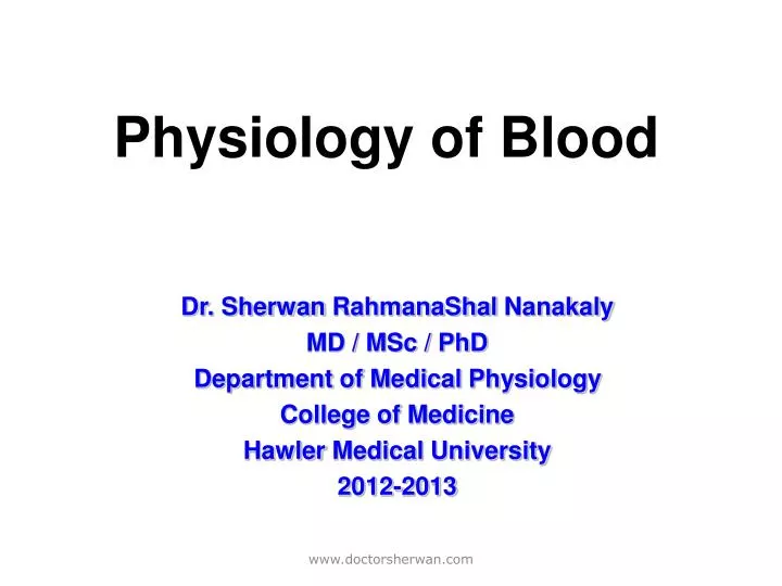
Physiology of Blood. Dr. Sherwan RahmanaShal Nanakaly MD / MSc / PhD Department of Medical Physiology College of Medicine Hawler Medical University 2012-2013. Outline. I. Blood composing II. Physical and chemical characteristics of blood III. Blood Cells

E N D
Physiology of Blood Dr. Sherwan RahmanaShalNanakaly MD / MSc / PhD Department of Medical Physiology College of Medicine Hawler Medical University 2012-2013 www.doctorsherwan.com
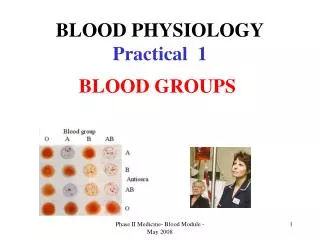
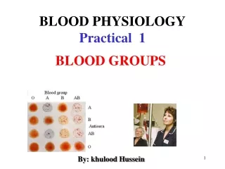
Outline I. Blood composing II. Physical and chemical characteristics of blood III. Blood Cells 1. Hemopoietic process and hemopoietic stem cells 2. Hemopoietic microenvironment 3. Erythrocyte Physiology 4. Leukocyte Physiology 5. Platelet or Thrombocyte Physiology IV. Physiological Hemostasis 1. Endocrine functions of vessel endothelial cells 2. Physiological Characteristics of Platelet 3. Blood Coagulation 4. Fibrinolysis V. Blood Group 1. RBC Agglutination 2. ABO blood group system 3. Rh blood group system 4. Relation between blood volume and clinic 5. Principle of Transfusion and Cross-match test General Objectives and Outline www.doctorsherwan.com
General Functions of the Blood • Transport: A: Gases; Oxygen, CO2, … B: Nutrient and Waste: Glucose, other carbohydrates, aminoacids….urea, creatinine, uric acid. C: Hormones and Enzymes. D: Antibodies. E: Electrolytes. www.doctorsherwan.com
General Functions of Blood , cont. II: Regulation: A: Temperature Regulation; warm blood (37-38 Co ) increased flow in cold exposure. B: pH Regulation: By buffer systems found in blood (Bicarbonate) that minimize chnages in pH. (Regulated between 7.35 to 7.45). C: Electrolyte Regulation (Na, Ka, Cl….). D: Blood pressure regulation: increasing and decreasing blood flow to the kidneys. www.doctorsherwan.com
General Functions of Blood , cont. III: Defense Mechanism: First line (Neutrophils) second line (Monocyte and Macrophages) via antibodies and third line (Complement Systems) all found in blood. IV: Miscellaneous : A: Homeostatic Mechanism: blood contains materials that stop bleeding (Hemostasis) this arrest of bleeding is part of Homeostasis (Stable Internal Environment). www.doctorsherwan.com
Blood composing • Blood composing: plasma + blood cells • Hematocrit: blood cells occupies the percentage of total blood volume. normal value male: 40-50% female: 37-48% newborn: 55% www.doctorsherwan.com
Chemical component of plasma • Water: > 90% • Small molecule: 2%, it is electrolytes, nutriment, metabolic products, hormone, enzyme, etc. • Protein: 60-80 g/L, plasma protein include albumin (40-50 g/L), globulin (20-30 g/L,α1-, α2, β-, γ- ) and fibrinogen. • Most of albumin and globulin made from liver. www.doctorsherwan.com
Function of plasma protein: • Transportation. • Nutrition. • Forming colloid osmotic pressure. • Coagulation and anticoagulation. • pH value buffer. • Immunity (globulin) www.doctorsherwan.com
Physical Characteristics of Blood • Average volume of blood: • 5–6 L for males; 4–5 L for females (Normovolemia) • Viscosity (thickness) - 4 - 5 (where water = 1) • The pH of blood is 7.35–7.45; x = 7.4 • Osmolarity = 300 mOsm or 0.3 Osm • This value reflects the concentration of solutes in the plasma • Salinity = 0.85% • Reflects the concentration of NaCl in the blood. www.doctorsherwan.com
Physical Characteristics of Blood 6. Temperature is 38C, slightly higher than “normal” body temperature. 7.Blood accounts for approximately 8% of body weight. 8. Specific gravity: total blood (1.050-1.060) more influenced by red blood cells; plasma (1.025-1.030) more influenced by plasma protein; RBC (1.090-1.092) more influenced by Hb www.doctorsherwan.com
Formed Elements (Blood Cells) Blood cells are erythrocyte (red blood cell, RBC), leukocyte (white blood cell, WBC) and thrombocyte (platelet, P). www.doctorsherwan.com
Blood Cells • The forming processes of erythrocyte (red blood cell, RBC), leukocyte (white blood cell, WBC) and thrombocyte (platelet, P) originating from hematopoietic stem cells are hemopoiesis. • Transfer of blood cells forming place: yolk sac hemopoiesis(earlyembryo period) →liver and spleen (second embryo month) → marrow↑and liver, spleen↓ (after fourthembryo month) → marrow (fetus birth time) and liver, spleen as complementary role. During adulthood (after 18), red marrow (flat bones, e.g. vertebra,ilium, sternum, rib, skull and long bone ending) rather than yellow marrow has hematopoietic functions. www.doctorsherwan.com
Erythrocyte Physiology Shape and number of red blood cells (RBC) • Shape of RBC: like biconcave disc Its diameter is about 7~8 µm, peripheral thickness about 2.5 µm, central thickness about 1 µm and cubage about 90 µm3. www.doctorsherwan.com
biconcave disc like Reason for shape of RBC www.doctorsherwan.com
Erythrocyte Physiology Number of RBC: It is most numbers in the blood. Normal value about RBC Male adult, 4.5~5.5×10¹²/L; average, 5.0×10¹²/L Female adult, 3.8~4.6× 10¹²/L; average, 4.2×10¹²/L Newborn, ≥ 6.0×10¹²/L Protein within RBC is hemoglobin (Hb). Hb in male adult, 120~160 g/L; Hb in female adult, 110~150 g/L; Hb in newborn (within 5 days), ≥ 200 g/L Pregnant female, numbers of RBC and Hb are relatively less (because of more plasma). www.doctorsherwan.com
Physiological Characteristics and Functions of RBC Characteristics of RBC 1. PERMEABILITY: semipermeable membrane, gas and urea freely passing through, negative ions easily in or out of RBC, and positive ions not. There are Na-K ATPase as pump on the membrane of RBC. 2. PLASTICITY AND METAMORPHOSE: Plasticity and metamorphose depend on: 1) surface area-cubage ratio, 2) viscosity of Hb, 3) membrane elasticity and viscosity. www.doctorsherwan.com
Physiological Characteristics and Functions of RBC Characteristics of RBC 3. Suspension stability: it cab be described by erythrocyte sedimentation rate (ESR) which is RBC descending distance per hour and suspension stability is inverse proportion to ESR. Normal value of ESR: male, 0~15 mm/h; female, 0~20 mm/1st h. ESR and clinic: some diseases bring about rouleaux formation (mainly involved in plasma component, e.g. globulin, fibrinogen, cholesterol) and speed up ESR. www.doctorsherwan.com
Physiological Characteristics and Functions of RBC Characteristics of RBC 4. Osmotic fragility: Changes in RBC put into lower osmotic salty solution. Osmotic fragility of aged RBC is large and easily results in rupture (hemolysis and ghost cell). Isosmotic solution, e.g. 0.85% NaCl, 1.4%NaHCO3, 5% glucose, etc. Isotonic solution, e.g. 0.85% NaCl Isosmotic solution does not equal to isotonic solution. Isosmotic solution, isotonic solution and clinic www.doctorsherwan.com
Physiological Characteristics and Functions of RBC Functions of RBC • RBC can be used for transportation of O2 and CO2 in the blood. • RBC can be served as pH buffer. www.doctorsherwan.com
Regulation of Erythropoiesis • 0.8% of total RBCs has self renewal, that is to say, 160×106 RBC production every minute. • Burst forming unit-erythroid, BUF-E, important to earlier erythropoiesis, depends on stimulation of burst promoting activity, BPA outside body. BPA made by leucocyte is a glycoprotein whose molecular weight is about 25000~40000 • Colony forming unit-erythroid, CFU-E, important to terminal erythropoiesis, depends on erythropoietin, EPO which is also a glycoprotein, molecular weight, 34000, plasma concentration 10 pmol/L, half life 5 hours, increasing release when anoxia. www.doctorsherwan.com
Life and breakage of RBC • Life-span: 120 days, about 4 months, each RBC circulates 27 km averagely in vessels, short life-span for aged RBC • Breakage: places are liver, spleen and lymphatic node, and after breakage, Hb released from RBC immediately combine with plasma α2-globulin (Hb touched protein) which is taken in by liver for iron reuse. • Hb, very toxic if it get into blood, normally, it can be metabolized into bile pigment in liver. www.doctorsherwan.com
Leukocyte PhysiologyClassification and numbers of Leukocyte • Number of Leukocyte (white blood cells, WBC): (4.0~10)×109/L • Classification: It is granulocyte (neutrophil, eosinophil, basophil), monocyte and lymphocyte. www.doctorsherwan.com
Classification and numbers of Leukocyte TABLE. Classification and normal value of Leukocyte Absolute Value (×109/L) Percentage (%) Total numbers of leukocytes 4.0~10.0 Neutrophil (bacilliform nucleus) 0.04~0.5 1~5 Neutrophil (foliiform nucleus) 2.0~7.0 50~70 Eosinophil 0.02~0.5 0.5~5 Basophil 0.0~0.1 0~1 Monocyte 0.12~0.8 3~8 Lymphocyte 0.8~4.0 20~40 For Clinic Use www.doctorsherwan.com
Physiological Changes in Numbers of Leukocyte • Newborn: Number is higher, 15×109/L, after birth 3 or 4 days to 3 months, being about 10×109/L, mainly, neutrophil, 70%; secondarily, lymphocyte. • Circadian changes: Number of WBC is more in the afternoon than in the morning. • Food taking, ache and mood excitation: Number of WBC is remarkably higher. • Heavy exercise and laboring: Increasing numbers, about 35×109/L, return to original level after action stop. • Terminal pregnancy of female: Numbers changes in 12~17×109/L, and during parturition, 34×109/L, and after parturition 2~5 days, number return to original level. www.doctorsherwan.com
WBC Diapedisis BloodVessel Chemotaxis Metamorphose Physiological Characteristics and Functions of WBC Terminology • Diapedisis: Metamorphosed WBCs pass through vessel wall getting into interstitial fluid. • Chemotaxis: It is a process that WBCs shift to some chemical material (metabolic production, antigen-antibody complex, bacteria, toxin, etc). • Phagocytosis: It is a process that WBCs enclose and engulf exotic or extraneous material, and use intracellular enzyme digesting them. www.doctorsherwan.com
Physiological Characteristics and Functions of WBC • Neutrophil • Another name, polymorphonuclear, PMN, 6~8 h in the vessels, diapedisis, chemotaxis and phagocytosis (using its hydrolyzed enzyme) • Function: It plays a very important role in nonspecific cellular immunity system which is against pathogenic microorganism, such as bacteria, virus, parasite, etc. • Clinic relation: Number of neutrophil greatly increase occurring in acute inflammation and earlier time of chronic inflammation. number decrease of neutrophil will result in poor resistibility and easily suffering from infection. www.doctorsherwan.com
Physiological Characteristics and Functions of WBC • Eosinophil • Circadian changes: Its number is lower in the morning and higher at night. • Function: 1. It limits and modulates the effects of basophil on fast allergic reaction. 2. It is involved in immune reaction against worm with opsonization. • Clinic relation: Its number increase when person suffers from parasite infection or allergic reaction. www.doctorsherwan.com
Physiological Characteristics and Functions of WBC • Basophil • Circulatory time: 12 hours • Basogranules contain heparin, histamine, chemotactic factors and chronic reactive material for allergic reaction. • Function: It is also involved in allergic reaction. 1. Heparin serves as lipase cobase and speeds up fatty decomposition. 2. Histamine and chronic reactive material increase permeability of capillary and contract bronchia smooth muscle, and result in allergic reaction such as measles, asthma. 3. Eosinophil chemotactic factor A released by basophil can attract eosinophil collection and modify eosinophil function. www.doctorsherwan.com
Physiological Characteristicsand Functions of WBC • Monocyte • Its body is large, diameter about 15~30 µm withoutgranule • Function: • 1. It contains many nonspecific lipase and displays the • powerful phagocytosis. • 2. As soon as monocytes get into tissue from blood , it change • name called macrophage activating monocyte- macrophage • system to release many cytokins, such as colony stimulating • factor (CSF), IL-1, IL-3, IL-6, TNFα, INF-α,β ,etc. • 3. Cytokins induced by monocyte may modulate other cells • growth. • 4. Monocyte- macrophage system plays a very important role in • specific immune responsive induction and regulation. www.doctorsherwan.com
Physiological Characteristicsand Functions of WBC • Lymphocyte • Classification: It can be separated into T- Lymphocyte and B- Lymphocyte. • Function: 1. Lymphocytes serve as a nuclear role in immune responsive reaction. 2. T- Lymphocytes involved in cellular immunity. 3. B- Lymphocytes involved in humoral immunity. • Clinic relation: Numbers increase of lymphocytes occur in chronic inflammation and late time of infection. www.doctorsherwan.com
Leukopoiesis, Regulation and Breakage • Birth place: bone marrow, originating from hemopoietic stem cells, and leukopoiesis process is similar to RBC. • Leukopoiesis, differentiation and growth are influenced by hemopoietic growth factor, HGF which are glycoprotein secreted by lymphocyte, monocyte- macrophage, fibrous cell and endothelial cell. • Colony stimulating factor, CSF, such as GM-CSF, G-CSF, M-CSF, Multi-CSF (IL-3) also influence Leukopoiesis. • Life span: several hours to 3 or 4 days. • Leukocyte breakage: site are liver, spleen and lymphatic node. • Pus or purulence forming www.doctorsherwan.com
Platelet or Thrombocyte Physiology • Shape: Biconvex disk like, diameter about 2~4 µm, average cubage 8 µm3. • Complicated structure: under the electronic microscope, there are α-granule, dense body, lysinperoxide enzyme, opening tubular system, dense tubular system, canaliculus,etc. • Dense body: It contains ADP, ATP, 5-HT, Ca2+, epinephrine,etc. • Source: Platelet comes from megakaryocyte fractionlet release in the marrow. www.doctorsherwan.com
Normal Value and Function of Platelet • Normal value: 100×109 ~ 300×109, range from 6%~10% • Normal changes: more number in the afternoon than in the morning, more in winter than in spring, more in the venous blood than capillary, after sport↑, pregnacy↑. • *Functions: 1. It maintains capillary endothelial cells smooth and integrated (repairing endothelium and providing nutrition). 2. It is involved in physiological hemostasis. • Platelet and clinic relation: decrease of platelet, abnormal immune reaction, will results in hemorrhage or bleeding, purpuric symptom. www.doctorsherwan.com
Life- Span and Breakage of Platelet • Life-span: Averagely, 7~14 days in the blood. It can be consumed when it displays physiological functions. • Breakage: Aged platelet can be processed by phagocytosis in liver, spleen and lymphatic node. www.doctorsherwan.com
Physiological Hemostasis • *Definition: The process from vessel bleeding to automatic hemostasia. • *Bleeding time: The time from vessel bleeding to automatic hemostasia. Normal time is 1~3 min and it is longer when platelet decrease. • Process of hemostasis: 1. Blood vessel contraction or convulsion (induced by neuroreflex; 5-hydroxytryptamine,5-HT; thromboxane A2, TXA2; endothelin, ET ) 2. Platelet thrombosis forming (made by platelet adhesion, aggregation, release and contraction) 3. fibrin, clot forming and maintenance (made by blood coagulation activation) www.doctorsherwan.com
Physiological Hemostasis www.doctorsherwan.com
Endocrine functions of vessel endothelial cells • Material related to hemostasis are basal membrane, collagen (III, IV), microfibril, elastin, laminin, ectonectin, fibronectin, von Willebrand factor (vWF), protein enzyme, protein enzyme inhibitor, adhesive amylose, etc. • Anticoagulative material: They are prostacyclin (PGI2), endothelium-derived relaxing factor (EDRF or nitric oxide, NO), tissue-type plasminogen activator (tPA), uPA, ADPase, ATIII, heparin sulfate, protein C, thrombomomodulin (TM), plasminogen activator (PA). • Promoting coagulative material: Tissue factor, vWF, blood clotting factor V, plasminogen activator inhibitor (PAI-1, PAI-2, ATIII), TNFα, interleukin-1 (IL-1). • Vessel constricting and relaxing modulators: endothelin-1 (ET-1), EDRF (NO), PGI2, etc. www.doctorsherwan.com
Roles of Vessel Endothelial Cells in Physiological Hemostasis Roles are close related to its endocrine functions • Vessel endothelium serves as barrier between underendothelial structure (namely, collagen) and blood. As soon as collagen expose to blood, hemostasis of platelet is immediately activated to form thrombus blocking wounded vessels. • Platelet activation can releases constrictive factors (TXA2, ET-1, 5-HT, etc) making vessel convulsion, lasting about 60 sec. • Stimulated vessel endothelial cells release coagulative factors and Promoting coagulative material to realize, speed up blood coagulation. At the same time, cells also release anticoagulative factors and fibrinolysis material to modify blood coagulation. www.doctorsherwan.com
Inactive Platelet Under the electronic microscope www.doctorsherwan.com
Activated Platelet for Hemostasis Under the electronic microscope www.doctorsherwan.com
Physiological Characteristics of Platelet • Thrombocyte adhesion:its membrane glycoprotein (GP,GPIb/IXand GPIIa/IIIb), collagen (underendothelial structure),vWF(plasma component),fibrinogen are involved in adhesion. Mechanism: Exposed collagen+vWF →vWF changes→platelet membrane glycoprotein+changed vWF → Thrombocyte adhesion. • Thrombocyte aggregation:induced by physiological factors such as ADP, thromboxane A2 (TXA2), epinephrine, 5-HT, histamine, collagen, thrombin, prostacyclin,etc and by pathological factors like bacteria, virus, immune complex, drugs, etc. The process can be separated into two phases: phase one is reversible aggregation and phase two irreversible aggregation. Two phases require Ca2+, fibrinogen and energy consumption. Mechanism : Various factors+corresponding receptors on the platelet →changes in the second messenger within platelet →cAMP↓, Ip3↑, Ca2+↑, cGMP↑→ platelet aggregation. www.doctorsherwan.com
Physiological Characteristics of Platelet • Thrombocyte release: ADP, ATP, 5-HT, Ca2+ released from dense body, and β-platelet globin, PF4, vWF, fibrinogen, PFV, PDGF, thrombin sensitive protein from α-granule, and acid protein hydrolyzed enzyme, tissue hydrolyzed enzyme from lysosome. • Thrombocyte contraction: Loose platelet thrombus could turn into compact platelet thrombus by Ca2+ release and cytoskeleton movement (filament/canaliculus) within platelet www.doctorsherwan.com
Roles of Platelet in Hemostasis • Activation of platelet: Stimulus brings about thrombocyte adhesion, aggregation, release and contraction. • Loose platelet thrombus forming: First phase of hemostasis. • Blood coagulation activation by platelet: Fibrin net forming, second phase of hemostasis. • *Roles of platelet in hemostasis: 1. Activated platelets supply lecithoid (phospholipid) surface for blood clotting factor and involve in activating factor X and prothrombin. 2. Surface of platelet membrane combine with many blood clotting factor, such as fibrinogen, FV, FXI, FXIII to speed up coagulation. 3. Activated platelets release α-granule which contains fibrinogen to intensify fibrin forming and blood coagulation. 4. Activated platelets contract clot with its contractive protein to solidify blood coagulation. www.doctorsherwan.com
Two Phases of PhysiologicalHemostasis First Phase Second Phase www.doctorsherwan.com
Mechanism1 of Platelet in Hemostasis www.doctorsherwan.com
Blood CoagulationBlood Clotting Factor • Definition: The process of blood flow from flowing liquid to gel or gelatin. • Serum: Light yellow fluid after blood coagulation. • Difference between serum and plasma mainly consists in no fibrinogen in serum. • Blood coagulation is a series of complicated biochemical reactions with various enzymes. • Blood clotting factor: Material which are directly involved in blood coagulation. There are 12 factors named Roman numerals, except Ca2+, phospholipid,other factors being protein, and except FIII (TF), others are in fresh plasma synthesized by liver with VitK . • Blood clotting enzymes have two type: inactive and activated type [FII, FVII, FIX, Fx, FXI, FXII, FXIII]. www.doctorsherwan.com
Blood Clotting Factor Factor Name Plasma Synthesizing Half life Chromsome Concentration site site I Fibrinogen 3000 Liver 4~5 d 4 II Prothrombin 100 Liver (with Vit K) 3 d 11 III Tissue factor - Endothelial cell - - IV Ca2+ 100 - - - V Proaccelerin 10 Endothelial cell, platelet 12~15 h 1 Ⅶ Proconvertin 0.5 Liver (with Vit K) 4~7 h 13 Ⅷ Antihemophilic factor,AHF 0.1 Liver 8~10 h Ⅹ Ⅸ Plasma thromboplastic 5 Liver (with Vit K) 24 h Ⅹ component,PTC(Christmas factor) Ⅹ Stuart-Prower Factor 10 Liver (with Vit K) 2 d 13 Ⅺ Plasma thromoboplastin 5 Liver 2~3 d 4 antecedent,PTA Ⅻ Contact factor or Hageman factor 40 Liver 24 h 5 XIII Fibrin-stabilizing factor 10 Liver, platelet 8 d 6,1 - High-molecular weight 80 Liver - 3 kininogen,HMW-K - Prekallikrein,Pre-K or Fletcher factor 35 Liver - 4 www.doctorsherwan.com
Blood Coagulation • Intrinsic pathway of blood coagulation: All blood clotting factors involved in blood coagulation come from blood. Eyewinker surface with negative charges (collagenin) on the endothelium of blood vessel activates blood FXII as beginning of coagulation named surface activation. • Extrinsic pathway of blood coagulation: Stimulus activates tissue factor (FIII) as beginning of coagulation. Extrinsic pathway of blood coagulation is faster than intrinsic pathway of blood coagulation because its steps are more simple. • *Basic steps of blood coagulation [typical positive feedback]: Prothrombin activator forming [FXa-Va-Ca2+-phospholipid] Step 1 Prothrombin thrombin Step 2 Fibrinogen fibrin (clot) Step 3 • Hemophilia A, B, C in the clinic results from deficiency of FVIII, FIX, FXI in the blood, respectively. www.doctorsherwan.com
Extrinsic pathway(Tissue Factor,TF) Intrinsic pathway( Eyewinker surface) Ⅻ TF+Ⅶ HK S Ⅺ Ca2+ K PK Ⅶ-TF Ⅸ Ⅻa Ⅹa Ca2+ Ca2+ ,PL Ⅺa Ⅶa-TF Ⅸa Ca2+ Ca2+ PL Ⅷa PL Ⅹ Ⅹa Ca2+ ⅩⅢ PL: phospholipid CL: cross linking fibrin HK: high molecular weight kininogen S: Subendothelium PK: prekallikrein K: kallikrein Ⅴa PL Ⅱ Ⅱa ⅩⅢa Ca2+ Ⅰ Ⅰa CL Ⅰa Process of Blood Coagulation www.doctorsherwan.com
Kallikrein (Intrinsic pathway) Endothelial cells (Extrinsic pathway) Cl-inhibitors (Urokinase, uPA) uPA uPAG tPA PAI-1 Plasminogen Plasmin α2-antiplasmin α2-huge globin Fibrin dissolution Fibrin or fibrinogen 4.Fibrinolysis • Fibrinolytic system is involved in fibrinolysis, tissue repair and vessel rebirth. • Two fibrinolytic systems: cellular one and plasma one. The former is leucocyte, macrophage, endothelial cell, mesothelial cell and platelet to engulf and digest fibrin. The latter is plasminogen activators (PA) and its inhibitors (PAI), plasminogen, plasmin. • Basic steps: www.doctorsherwan.com