Download
1 / 43
530 likes | 1.32k Vues
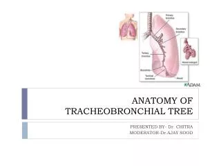
Anatomy Of Tracheo -Bronchial Tree. Dr. Supreet Singh N ayyar, AFMC f or more presentations, visit www.nayyarENT.com. Layout. Embryology Respiratory Tree Trachea and main bronchi Segmental Bronchi Bronchopulmonary Segments Structure Vasculature , Lymphatic and Nerve Supply

E N D
Anatomy Of Tracheo-Bronchial Tree Dr. Supreet Singh Nayyar, AFMC for more presentations, visit www.nayyarENT.com www.nayyarENT.com
www.nayyarENT.com Layout • Embryology • Respiratory Tree • Trachea and main bronchi • Segmental Bronchi • Bronchopulmonary Segments • Structure • Vasculature , Lymphatic and Nerve Supply • Applied Anatomy
www.nayyarENT.com EMBRYOLOGY • Tracheobronchial Diverticulum below Hypobranchial Eminence – 4th wk IUL • edges – oesophagotracheal septum • fuses caudally • endoderm lining • cranial – Trachea / Larynx • caudal – Bronchi / Lungs
www.nayyarENT.com EMBRYOLOGY
www.nayyarENT.com EMBRYOLOGY
www.nayyarENT.com The Respiratory Tree
www.nayyarENT.com The Trachea • Cartilaginous and Membranous tube • Dimensions • Length -10- 12 cm • Ext Dia – 2.0 cm ( 1.5 cm) • Extent – C6 – T4 • Incomplete rings
www.nayyarENT.com Relations • Cervical • Anteriorly • Isthmus of thyroid with inf thyroid vessels • Pretracheal fascia • Sternothyroid & Sternohyoid • Investing layer of Deep Cervical Fascia • Skin & superficial fascia • Posteriorly • Oesophagus; longus colli; RLN • On each side • Thyroid • Common Carotid Artery
Relation o cervical trachea at C 7 www.nayyarENT.com
www.nayyarENT.com Relations • Thoracic • Anterior • ManubriumSterni • Sternothyroid • Thymus • Lt Brachiocephalic V • Aorta & Lt CCA • Deep Cardiac Plexus • Posterior • Oesophagus • Vertebral Column • Rt Side • Right Lung & Pleura • RtVagus • Azygous V • Lt Side • Arch of Aorta • Lt Common Carotid • Lt Subclavian A • Lt RLN
www.nayyarENT.com The main Bronchi and Branches • Rt and Lt Main Bronchi – T4 level • Carina • The Right Bronchus (bronchus dexter), wider, shorter, and more vertical • 5 cm. long • Enters right lung opposite fifth thoracic vertebra • Azygos vein arches over it from behind • Right pulmonary artery lies at first below and then in front • 2.5 cm along course- Rt Upper Lobe Bronchus • At Hilum of Lung – Middle & Lower Lobe Bronchi
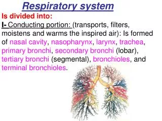
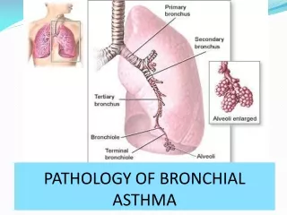
Bronchi, Bronchioles, & Alveoli www.nayyarENT.com • Tracheadivides into • Primary bronchiidivide into • Secondary bronchidivide into • Tertiary bronchidivide into • Bronchiolesdivide into • Terminal bronchiolesdivide into • Respiratory bronchioles divide into • Alveolar ductsend in • Alveoli
www.nayyarENT.com The Respiratory Tree
www.nayyarENT.com The Respiratory Tree Structure through the airways 1.Trachea 2. Bronchus (Right- or Left- Primary Bronchus) 3. Lobar Bronchus 4. Segmental Bronchus 5. Bronchus 6. Bronchiole 7. Terminal Bronchiole 8. Respiratory Bronchiole 9. Alveolar Duct 10. Alveolar Sac / Alveolus
www.nayyarENT.com The Respiratory Tree • Rt Upper Lobe Bronchus • Rt lat aspect of bronchus • Superolaterally • 3 segmental Bronchi ( Apical; Post; Ant ) • Apical Segmental – Apical & Ant. Subsegmental Br • Post Segmental – Lateral & Ant. Subsegmental Br • Ant. Segmental – Lateral & Ant. Subsegmental Br • Rt Middle Lobe Bronchus • 2.5 cm beyond Rt Upper Lobe Bronchus • Ant aspect • Forwards Downwards and Laterally • Lateral and Medial Subsegments
www.nayyarENT.com The Respiratory Tree • Rt lower lobe bronchus • Apical Segmental – post aspect Medial ; Sup ; Lat subsegmentalbr • Medial Basal ( Cardiac ) – Rt Border of heart • Ant Basal • Lateral Basal • Post Basal
www.nayyarENT.com The Respiratory Tree
www.nayyarENT.com The Respiratory Tree
www.nayyarENT.com The Respiratory Tree • Left Bronchus (bronchus sinister) smaller in calibre but longer than the right • 5.5 cm long • Enters root of the left lung opposite sixth thoracic vertebra • Passes beneath Aortic arch, • crosses in front of oesophagus, thoracic duct, descending aorta • Left pulmonary artery lies at first above then in front
www.nayyarENT.com The Respiratory Tree • Lt upper Lobe Bronchus • Anterolateral aspect 5.5cm from carina • Curves laterally then divides • Cranial divisions: Apico posterior and Anterior • Caudal divisions : anterolaterallyLingular area of lung Sup and Inf segmental • Lt Lower Lobe Bronchus • Apical segmental • Originates posteriorly 1cm below upper lobe orifice • Anteromedial (Anterior Basal and Medial Basal ) • PosterolateralLateral Basal and Post Basal)
The Lungs www.nayyarENT.com • Soft, spongy, cone-shaped • Right lung • 3 lobes • Left lung • 2 lobes
www.nayyarENT.com Bronchopulmonary segments • Independent respiratory units • Sectors of lung with tertiary/ segmental bronchus ;separate Br of Pulmonary A • Pyramidal with apex to root of lung • Clinical Significance • Restriction of infections • Segmental resection • Bronchoscopy
www.nayyarENT.com Structure of Trachea andMajor Bronchi • Cartilages • 16-20 • Incomplete Rings (4 mm vertically and 1 mm thick) • Union of 2 or more rings partially or completely • Highly elastic • 1st Tracheal Cartilage • Fibrous Membrane • Perichondrium of cartilages • Collgen with elastic fibres • Non striated musfibres
Changes as the Respiratory Tree Branches www.nayyarENT.com • As branching of bronchi becomes more extensive • Rings of cartilage become plates • Smooth muscle increases • Columnar epithelium becomes cuboidal, then squamous (in alveoli
www.nayyarENT.com • Mucous Membrane • Epith lining – thins and single layered • Tubulo-racemose glands • Not present in bronchioles
www.nayyarENT.com Vasculature and lymphatics • Blood Supply • Inf Thyroid A • Bronchial A – 1 for Rt lung ;2 for Lt • Lie against post wall of Bronchi • 2 Bronchial Veins • Rt – Azygous V • Lt – Lt Sup Intercostal V or HemiazygousV • Pulmonary V
www.nayyarENT.com Vasculature and lymphatics • Lymphatic Drainage • Pretracheal and Paratracheal groups • Superficial System- Tissue beneath pleura vessels round lung borders and fissures to reach hilum numerous Valves • Deep System- Bronchial Tree; Pulm Vessels hilum- drain into Bronchopulmonary Nodes less no. of Valves • Rt and Lt Paratracheal LN unite with Internal Thoracic and Brachiocephalic LN
www.nayyarENT.com Nerve Supply • Parasympathetic ( Vagus) • Motor to bronchial muscle – bronchospasm • Secretomotor to mucous gland • Sensory- stretch and Cough Reflex • Sympathetic • Cervical Ganglion and Spinal N. T2 – T5 • Inhibitory to smooth muscle and glands
www.nayyarENT.com Applied Anatomy • Tracheostomy • Bronchoscopy • Foreign Body In Respi Tract
www.nayyarENT.com Thank youfor more presentations, visit www.nayyarENT.com