Download
1 / 55
580 likes | 1.15k Vues
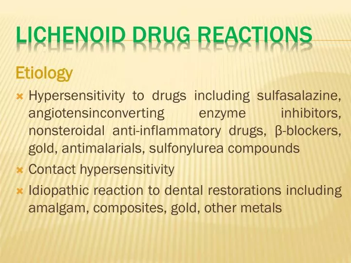
Etiology Hypersensitivity to drugs including sulfasalazine, angiotensinconverting enzyme inhibitors, nonsteroidal anti-inflammatory drugs, β-blockers, gold, antimalarials, sulfonylurea compounds Contact hypersensitivity

E N D
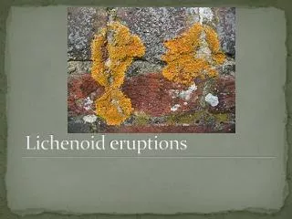
Etiology Hypersensitivity to drugs including sulfasalazine, angiotensinconverting enzyme inhibitors, nonsteroidal anti-inflammatory drugs, β-blockers, gold, antimalarials, sulfonylurea compounds Contact hypersensitivity Idiopathic reaction to dental restorations including amalgam, composites, gold, other metals Lichenoid Drug REACTIONS
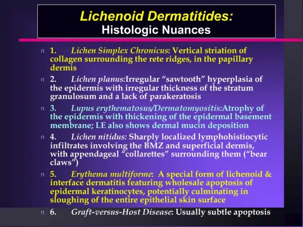
Clinical Presentation • White striae or papules, as with lichen planus • Lesions may appear ulcerative with associated tenderness or pain. • Most often in buccal mucosa and attached gingiva, but any site may be involved
Diagnosis • Identification and elimination of causative substance. • Biopsy of areas unresponsive to elimination strategy to demonstrate characteristic keratosis and interface inflammation and associated changes. • Patch testing performed to confirm contact allergens. Differential Diagnosis • Lichen planus • Leukoplakia • Dysplasia/carcinoma
Treatment • Alternative drugs or material to be chosen • Topical corticosteroid applications • Topical tacrolimus applications Prognosis • Good • Observation while lesions exist
Etiology • An autoimmune-/immunologically mediated condition • Antibodies demonstrable against an array of cytoplasmic and nuclear antigens • Most often occurs in women Lupus Erythematosus
Clinical Presentation • Three forms are as follows: • Chronic cutaneous (CCLE) or discoid (DLE) • Subacute cutaneous (SCLE) • Systemic (SLE) • Black females have highest incidence • Predominates in women over 40 years • 80% of patients have concurrent cutaneous findings • 30 to 40% of SLE patients have oral mucosal findings • Oral mucosal lesions may appear Lichenoid, keratotic, and erosive. • Labial vermilion with crusted, exfoliative, erythematous, and keratotic appearance • Oral findings are most common in CCLE or DLE.
Diagnosis • Direct immunofluorescent examination of mucosal biopsy • Serologic correlation (antinuclear antibodies: anti–SS-A, –SS-B, and double-stranded deoxyribonucleic acid) Differential Diagnosis • Lichen planus • Candidiasis • Hypersensitivity/lichenoid eruption • Leukoplakia
Treatment • Complex—dependent on LE variant present and level of disease expression • Systemic corticosteroids and immunosuppressive agents for SLE • Topical corticosteroid agents for intraoral lesions • Low-dose hydroxychloroquine • Intralesional corticosteroid injections Prognosis • Good prognosis with CCLE or DLE form • Variable prognosis with SLE • SCLE has an intermediate prognosis between that of SLE and CCLE or DLE forms.
Etiology • Ectopic sebaceous glands within the oral mucosa and vermilion portion of the lips Fordyce’s Granules
Clinical Presentation • Multiple, scattered, yellowish pink, maculopapular granules • Buccal mucosa and vermilion of lips predominantly affected • Asymptomatic • Increasingly prominent after puberty
Diagnosis • Bilateral distribution and appearance • Lack of symptoms • If biopsy performed, normal sebaceous glands in the absence of hair follicles noted Differential Diagnosis • Candidiasis Treatment • None • Reassurance Prognosis • Excellent
Etiology • Unknown; may be familial • May be related to atopy • Small percentage associated with cutaneous psoriasis Geographic Tongue
Clinical Presentation • May be symptomatic in association with spicy or acidic foods • Focal red depapillated areas bordered by slightly elevated, yellowish margin • Dynamic behavior: changes in shape, size, intensity day to day • Dorsal and lateral tongue surfaces affected predominantly • Ventral tongue and other areas less often involved • Often associated with fissured tongue
Diagnosis • Location and appearance • Biopsy confirmation usually unnecessary Differential Diagnosis • Reiter’s syndrome • Lichen planus • Lupus erythematosus • Candidiasis • Psoriasis
Treatment • None, if asymptomatic • Topical corticosteroids, if symptomatic Prognosis • Excellent • No malignant potential • May last months to years with periods of remission
Etiology • Generally unknown • May be related to poor oral hygiene, soft diet, heavy smoking, systemic or topical antibiotic therapy, radiation therapy, xerostomia, or use of oxygenating mouth rinses (H2O2, sodium perborate) Hairy Tongue
Clinical Presentation • Elongated, hyperkeratotic filiform papillae on tongue dorsum producing a “furred” to “hairy” texture • Color varies from tan to brownish yellow to black depending upon diet, drugs, chromogenic organisms • Symptoms usually minimal; may produce gagging or tickling sensation on palate
Diagnosis • Clinical features • Culture or cytologic studies not helpful Treatment • Physical débridement (brushing with a soft-bristled toothbrush, 5 to 15 strokes, once or twice daily) • Topical podophyllin (5% in benzoin) followed by débridement • Elimination of cause, if identified Prognosis • Excellent
Etiology • Results from direct mucosal contact with a quid containing areca (betel) nut, tobacco, and other ingredients; alkaloids and tannin in the areca nut are liberated by action of slaked lime within the quid, which is wrapped with the betel leaf • Risk of oral squamous cell carcinoma is increased several-fold Submucous Fibrosis
Clinical Presentation • Early phase: tenderness, vesicles, erythema, burning, melanosis • Later phase: mucosal rigidity, trismus • Sites most often affected: buccal mucosa, soft palate • Leukoplakia of surface with pallor • Deep scarring, epithelial atrophy in cheeks, soft palate
Microscopic Findings • Biopsy results show submucosal deposition of dense collagen. • Epithelial thinning, hyperkeratosis • Epithelial dysplasia found in up to 15% of cases Diagnosis • Appearance • History Differential Diagnosis • Lichen sclerosus
Treatment • Intralesional corticosteroid placement • Surgical release of scar bands in latter stages • Careful follow-up and vigilance for development of squamous cell carcinoma Prognosis • Irreversible • Fair
▼ BLUE/PURPLE VASCULAR LESIONS Hemangioma Varix Angiosarcoma Kaposi’s Sarcoma Hereditary Hemorrhagic Telangiectasia ▼ BROWN MELANOTIC LESIONS Ephelis and Oral Melanotic Macule Nevocellular Nevus and Blue Nevus Malignant Melanoma Drug-Induced Melanosis Physiologic Pigmentation Café au Lait Pigmentation Smoker’s Melanosis Pigmented Lichen Planus Endocrinopathic Pigmentation HIV Oral Melanosis Peutz-Jeghers Syndrome ▼ BROWN HEME-ASSOCIATED LESIONS Ecchymosis Petechia Hemochromatosis ▼ GRAY/BLACK PIGMENTATIONS Amalgam Tattoo Graphite Tattoo Hairy Tongue Pigmentation Related to Heavy-Metal Ingestion
Etiology • Benign developmental anomalies of blood vessels that may be subclassified as congenital hemangiomas and vascular malformations • “Congenital hemangioma” usually noted initially in infancy or childhood (hamartomatous proliferation) • Congenital hemangioma due to proliferation of endothelial cells • “Vascular malformations” due to abnormal morphogenesis of arterial and venous structures Hemangioma
Clinical Presentation • Congenital lesions usually arise around time of birth, grow rapidly, and usually involute over several years. • Malformations generally are persistent, grow with the child, and do not involute. • Color varies from red to blue depending on depth, degree of congestion, and caliber of vessels • Range in size from few millimeters to massive with disfigurement • Most common on lips, tongue, buccal mucosa • Usually asymptomatic • Sturge-Weber syndrome (trigeminal encephaloangiomatosis) includes cutaneous vascular malformations (port wine stains) along trigeminal nerve distribution, mental retardation, and seizures.
Diagnosis • Aspiration • Blanching under pressure (diascopy) • Imaging studies Differential Diagnosis • Purpura • Telangiectasia • Kaposi’s sarcoma • Other vascular neoplasms
Treatment • Observation • Congenital hemangiomas typically involute, whereas vascular malformations persist. • Surgery (scalpel, cryosurgery, laser [argon, copper])—congenital hemangiomas usually are circumscribed and more easily removed than are vascular malformations, which are poorly defined. (Vascular malformations are associated with excessive bleeding and recurrence.) • Sclerotherapy • Microembolization followed by resection for large malformations or if bleeding is problematic Prognosis • • Guarded
Etiology • An abnormal venous dilatation • Congenital or from damage to vessel wall (trauma, ultraviolet light) • Occur with increasing frequency over 40 years of age Varices
Clinical Presentation • Blue, lobulated surface • Painless, evolves slowly • Common on lower lip, sublingual regions • Blanches with compression (diascopy) • May become thrombosed
Diagnosis • Clinical appearance • Histological viewing of large-caliber, thin-walled vein Differential Diagnosis • Mucocele • Vascular neoplasm • Blue rubber bleb nevus syndrome • Hereditary hemorrhagic telangiectasia (Rendu-Osler-Weber syndrome)
Treatment • Observation only, if stable • Elimination by excision, sclerotherapy, or laser ablation Prognosis • Excellent
Etiology • Several forms • Classic idiopathic form affecting extremeties • Endemic form (African) • Immunosuppression-associated form • Acquired immunodeficiency syndrome associated form • All forms, especially AIDS-associated and immunosuppression associated forms, may be caused by or closely related to a herpesvirus (human herpesvirus 8 [HHV-8] or Kaposi’s sarcoma–associated herpesvirus [KSHV]). Kaposi’s Sarcoma
Clinical Presentation • Classic form associated with slow but pernicious growth over many years; oral lesions rarely seen • Endemic form more rapid; oral lesions rarely seen • AIDS-associated KS most commonly seen on keratinized mucosa/ mucoperiosteal tissues; strong predilection for hard palate, followed by gingiva, buccal mucosa, and tongue (prevalence decreasing with treatment for AIDS) • Evolution from bluish macule to nodule(s) • Evolution to multiple lesions • May precede or follow cutaneous lesions • Usually asymptomatic
Diagnosis • Location and appearance • May occur in up to one-third of AIDS patients • Biopsy showing spindle cell proliferation with vascular slits, extravascular red blood cells Differential Diagnosis • Hematoma • Hemangioma • Ecchymosis • Malignant melanoma • Pyogenic granuloma • Amalgam tattoo
Treatment of AIDS-Associated Form Radiation therapy: single fraction or equivalent fractionated therapy of 800 cGy Intralesional therapy: interferon-α, vincristine, vinblastine (2 mg/cc), sclerosing agents (sodium morrhuate) Systemic chemotherapy: interferon-α, vincristine, vinblastine, bleomycin, daunorubicin Most treatment is palliatively directed. Prognosis Variable, depending upon host’s immune status, but generally poor in AIDS-associated form
Angiosarcoma is a rare neoplasm of endothelial cell origin. A distinctive clinical pathologic variant of angiosarcoma is Kaposi's sarcoma. • The scalp is the usual location for angiosarcomas, although occasional lesions have been reported in the maxillary sinus and oral cavity. • The lesion consists of an unencapsulated proliferation of anaplastic endothelial cells enclosing irregular luminal spaces. • It has an aggressive clinical course and a poor prognosis. Angiosarcoma