Download
1 / 90
950 likes | 1.36k Vues
ACC/AHA 2005 Practice Guidelines for the Management of Pts With Peripheral Arterial Disease (Lower Extremity). peripheral arterial disease. Encompasses a large series of disorders affecting arterial beds exclusive of the coronary arteries. LOWER EXTREMITY PAD-Risk Factors. Cigarette smoking.

E N D
ACC/AHA 2005 Practice Guidelines for the Management ofPts With Peripheral Arterial Disease (Lower Extremity)
peripheral arterial disease • Encompasses a large series of disorders affecting arterial beds exclusive of the coronary arteries
Cigarette smoking • 2 to 3 times more likely to cause lower extremity PAD than CAD • Increases the risk of lower extremity PAD by 2- to 6-fold and the risk of intermittent claudication by 3- to 10-fold • More than 80% of patients with lower extremity PAD are current or former smokers
Diabetes mellitus • increases the risk of lower extremity PAD by 2- to 4-fold • present in 12% to 20% of persons with lower extremity PAD
classic IC • pain, ache, tightening, cramping, or sense of fatigue in one or more of the lower extremity muscle groups • triggered by ambulation & relieved by rest • have sufficient blood flow so that limb ischemic symptoms are absent at rest. • site of claudication is distal to the diseased arterial segment • buttock, hip, and thigh claudication are seen with aortoiliac disease and calf claudication with femoral-popliteal disease
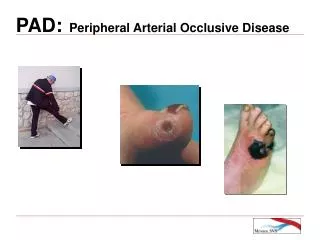
critical limb ischemia • rest pain, cold, or numbness of the feet, with or without tissue loss (nonhealing ulcers or gangrene). • Rest pain usually occurs at night (because of the horizontal position, which deprives the patient of the effect of gravity on blood flow through the tight lesions) • improves when the legs are in the dependent position. • superimposed edema of the affected leg(s) occasionally may be seen in those who tend to dangle their legs overnight.
Foot Physical Examination and Differential Diagnosis of Neuropathic and Neuroischemic Ulcers
Risk of Cardiovascular Events • 20% to 60% increased risk for MI • 2- to 6-fold increased risk of death due to CAD • Risk of stroke is increased by approximately 40% • Men with LL-PAD -4 to 5 times more likely to have a stroke / TIA
Ankle-Brachial Index and Severity of Peripheral Arterial Disease
Acute limb ischemia • rapid or sudden decrease in limb perfusion • threatens tissue viability • form of CLI, may be 1stmanifestation of arterial disease in asymptomatic pt • form of CLI, may occur as an acute event causing symptomatic deterioration in a pt with antecedent LL-PAD and IC • Progression of PAD from IC to CLI – gradual (may also reflect cumulative effect of multiple acute local thrombotic events that progressively increase the intensity of ischemia)
Paresthesia and paralysis imply irreversible ischemia, and muscle rigidity is a sign of a nonsalvageable limb.
Society for VascularSurgery/International Society for Cardiac Vascular Surgery(SVS/ISCVS)
Magnetic Resonance Angiography • RECOMMENDATIONS Class I • 1. MRA of LL -diagnose anatomic location and degree of stenosis of PAD ( A) • 2. MRA of LL should be performed with gadolinium enhancement ( B) • 3. MRA of LL -useful in selecting pts with LL-PAD as candidates for endovascular intervn( A) Class IIb • 1. MRA of LL -may be considered to select pts with LL-PAD as candidates for surgical bypass and to select the sites of surgical anastomosis. (B) • 2. MRA of LL -may be considered for postrevascularization surveillance in pts with LL-PAD. (B)
MRA limitations • Tends to overestimate degree of stenosis because of turbulence • May overestimate occlusions owing to loss of signal from retrograde collateral flow • Metal clips can cause artifacts that mimic vessel occlusions • Some metal stents will obscure vascular flow • Pts with PPI and ICD and some cerebral aneurysm clips cannot be scanned safely • MRA performed with gadolinium has on rare occasions been associated with renal toxicity in patients with elevated creatinine levels
Computed Tomographic Angiography • RECOMMENDATIONS Class IIb • 1. CTA of LL may be considered -anatomic location and presence of signi stenosis in pts with LL-PAD (B) • 2. CTA of LL may be considered as a substitute for MRA for pts with CI to MRA(B)
CTA has potential advantages over MRA • Pts with PPI or ICD may be imaged safely with CTA • Metal clips, stents, and prostheses usually do not cause significant CTA artifacts • Has higher resolution • Can provide images of calcification in the vessel wall • Scan times are significantly faster with CTA than with MRA • Claustrophobia not a problem CTA also has potential disadvantages compared with MRA • Requires iodinated contrast, which may be nephrotoxic • Requires ionizing radiation
PAG-RECOMMENDATIONS Class I • 1. Recommended for evaluation of patients with LL-PAD when revascularization is contemplated. (B) • 2. Signi of an obstructive lesion is ambiguous → trans-stenotic P-gradients & supplementary angulated views to be obtained. (B) • 3. Pts with baseline renal insufficiency should receive prior hydration. (B) • 4. Follow-up clinical evaluation (physical Ex & RFT)-recommended ≤2 weeks after contrast angio to detect presence of delayed adverse effects, such as atheroembolism, ↓ in RFT, or access site injury (C)
Class IIa • 1. Noninvasive imaging modalities, including MRA, CTA, and color flow duplex imaging, may be used in advance of invasive imaging to develop an individualized diagnostic strategic plan, including • assistance in selection of access sites, • identification of significant lesions, and • determination of the need for invasive evaluation • 2. Treatment with n-acetylcysteine in advance of contrast angiography is suggested for pts with baseline renal insufficiency (Cr>2.0 mg/Dl)
CILOSTAZOL-RECOMMENDATIONS Class I • 1. Cilostazol (100 mg BD) is indicated to improve symptoms & ↑walking distance in pts with LL-PAD & IC (in the absence of CCF). (A) • 2. A therapeutic trial of cilostazol should be considered in all patients with lifestyle-limiting claudication (in the absence of CCF). (A)
PENTOXIFYLLINE-RECOMMENDATIONS Class IIb • 1. Pentoxifylline (400 mg TID) 2-line alternative therapy to cilostazol to improve walking distance in patients with IC (A) • 2. The clinical effectiveness of pentoxifylline as therapy for IC is marginal & not well established.(C)
Lipid-Lowering Drugs-RECOMMENDATIONS Class I • Statins indicated for all pts with PAD to achieve a target LDL < 100 mg/Dl Class IIa • 1. Statins to achieve a target LDL <70 mg/dL is reasonable for pts with LL-PAD at very high risk of ischemic events • 2. Fibrates can be useful for pts with PAD and low HDL , normal LDL, & elevated TGL
Antiplatelet and Antithrombotic Drugs RECOMMENDATIONS Class I • 1. Antiplatelets indicated to ↓ risk of MI, stroke, or vascular death in pts with atherosclr LL-PAD • 2. Aspirin ( 75 - 325 mg)- safe & effective antiplatelet therapy • 3. Clopidogrel (75 mg ) -effective alternative antiplatelet therapy to aspirin Class III • Oral anticoagulation therapy with warfarin is not indicated
Indications for Revascularization in IC Before a pt with IC is offered any invasive therapy,thefollowing considerations must be taken into account: • a predicted or observed lack of adequate response to lifestyle therapy and pharmacotherapies • the presence of a severe disability • absence of other disease that would limit exercise even if IC improved • the morphology of the lesion (appropriate intervention would have ↓risk & a ↑ probability of initial & long-term success)
Morphological Stratification of Iliac Lesions TASC type A iliaclesions: • 1. Single stenosis <3 cm of the CIA or EIA (unilateral/bilateral) TASC type B iliaclesions: • 2. Single stenosis 3 to 10 cm in length, not extending into the CFA • 3. Total 2 stenoses <5 cm in CIA and/or EIA & not extending into CFA • 4. U/L CIA occlusion TASC type C iliaclesions: • 5. B/L 5-10 cm-long stenosis of CIA and/or EIA, not extending into CFA • 6. U/L EIA occlusion not extending into the CFA • 7. U/L EIA stenosis extending into the CFA • 8. B/L CIA occlusion TASC type D iliaclesions: • 9. Diffuse, multiple U/L stenoses of CIA, EIA, & CFA (usually >10 cm) • 10. U/L occlusion involving both the CIA and EIA • 11. B/L EIA occlusions • 12. Diffuse disease involving Ao& both iliac arteries • 13. Iliac stenoses with an abdAoaneu/other lesion requiring Ao/iliac sx
Morphological Stratification of Femoropopliteal Lesions TASC type A femoropopliteal lesions: • 1. Single stenosis <3 cm SFA or popliteal A TASC type B femoropopliteal lesions: • 2. Single stenosis 3 - 10 cm, not involving distal popliteal A • 3. Heavily calcified stenoses ≤3 cm in length • 4. Multiple lesions, each <3 cm (stenoses or occlusions) TASC type C femoropopliteal lesions: • 6. Single stenosis or occlusion >5 cm • 7. Multi stenoses or occlusions, each 3-5 cm, with or without heavy calcification TASC type D femoropopliteal lesions: • 8. Complete CFA or SFA occlusions or complete popliteal and proximal trifurcation occlusions
Endovascular Treatment for IC- RECOMMENDATIONS Class I • Preferred revascularization technique for TASC type A iliac & femoropopliteal lesions • Translesional P gradients (with and without vasodilation) should be obtained to evaluate signi of angiographic iliac stenoses of 50%-75% D before intervn • Provisional stentng indicated for use in iliacs as salvage therapy for a suboptimal/failed result from POBA Class IIa • Stents can be useful in femoral, popliteal, & tibial arteries as salvage for a suboptimal or failed result from POBA
Summary of preferred options in interventional management of iliac lesions
Summary of preferred options for interventional treatment of femoropopliteal lesions
Thrombolysis for A/c & C/C LI -RECOMMENDATIONS Class I • Catheter-based thrombolysis is indicated for pts with a/c limb ischemia of ≤14 days (A) Class IIa • Mechanical thrombectomy devices can be used as adjunctive therapy for a/c LI due to peripheral arterial occlusion
ACC/AHA 2005 Practice Guidelines for the Management ofPts With Peripheral Arterial Disease (Renal artery)
Clinical clues to RAS • Onset of HTN < 30 yrs (I) • Severe HTN >55 yrs (I) • Accelerated, resistant or malignant HTN (I) • New azotemia/wors RFT aft adm of ACEI/ARB (I) • Unexpl atrophic kidney/size discrep >1.5 cm (I) • Sudden unexplPulm Edema (I) • Unexpl ↓RFT, inc pts on RRT (IIa) • Multi-vessel CAD (IIb) • Unexplained CCF (IIb) • Refractory angina (IIb)
Prevalence in gen population (RA duplex) • ≥65 yrs (834) • →6.8% (Women-5.5% , Men-9.1%) Screening RA angio at CAG • RA disease →30% • Significant RAS→11%-18% • Significant RAS →22%-59% pts with PAD ESRD in HD >20-years (683pts) • 12% had documented RAS as a cause of ESRD.
Renal function in pts with atheroRAS on medical therapy (28/12 follow up) • 46%→ ↑S.Cr • 29%→ 25%-50% ↓GFR • 37%→↓kidney size by > 10%
Median survival for • ESRD with renovasculardisease- 25/12 • ESRD due to malignant HTN- 55/12 • ESRD due to polycystic KD- 133/12
Clinical Clues to the Diagnosis of RAS –RECOMMENDATIONSPerformance of diagnostic studies Class I • 1. Onset of HTN <30 yrs • 2. Onset of severe HTN >55 yrs • 3. Characteristics: • (a) acc HTN (sudden & persistent worsening of prev controlled HTN) • (b) resistant HTN • (c)malign HTN (A/C decompCCF, A/C visual/neurodisturb &/ adv retinopathy) • 4. New azotemia /Worsening RFT aft an ACEI or an ARB • 5. Unexpatrophic kidney/Discrepancy in size of >1.5 cm • 6. Sudden, unexppulmedema (espazotemic pts) Class IIa • 1.Unexp RF, including pts starting RRT
Class IIb • 1. The performance of arteriography to identify signi RAS - reasonable in pts with multivessel CAD & none of the clinical clues or PAD at the time of arteriography.(B) • 2. Diagnostic studies -reasonable in pts with unexpl CCF or refractory angina(C)
Atherosclerosis • 90% of all renovascularstenotic lesions • Most often affects the aorto-ostial segment, including the proximal 1 cm of main RA
Fibromuscular Dysplasia • nonatherosclerotic, noninflammatory disease • HTN in a young woman (can occur in both genders at any age) • Middle & distal ⅔ of main RA • May involve RA branches (25%- segmental arteries) • Tends to occur in 25 - 50 yr old women • Often involves both RA (B/L in 60% ) • Characteristic - “string of beads” appearance • Medial fibroplasia≈ 80% of FMD • Intimalfibroplasia- relatively rare (thin, discrete web)
Also affects other arteries, inclu Carotid & Vertebral, & less commonly, Iliac & Mesenteric • Appears to be an assobetw Carotid & Vertebral FMD and intracranial aneurysmal disease • MRA of head should be performed in all patients with cervicocranial FMD
RenovascularHTN may also be caused by renal artery aneurysms • Aneurysm rupture is of greatest concern with noncalcifiedaneurysms >2 cm D, particularly in premenopausal women
Other causes of renovascular disease • Takayasu’sarteritis • Atheroemboli • Thromboemboli • William’s syndrome • Neurofibromatosis • Spontaneous renal artery dissection • Arteriovenousmalformations or fistulas • Trauma (e.g.,lithotripsy, direct injury, or surgery) • Prior abdominal radiation therapy • Retroperitoneal fibrosis

![Diffuse Vascular Disease (Focus on Peripheral Arterial Disease [PAD])](https://cdn1.slideserve.com/3363465/slide1-dt.jpg)