The Cell Membrane
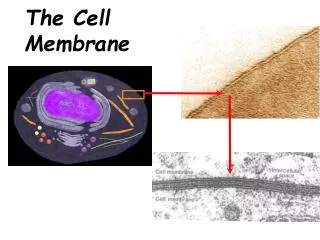
The Cell Membrane . Overview. Cell membrane separates living cell from nonliving surroundings thin barrier = 8nm thick Controls traffic in & out of the cell selectively permeable allows some substances to cross more easily than others hydrophobic vs hydrophilic

The Cell Membrane
E N D
Presentation Transcript
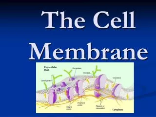
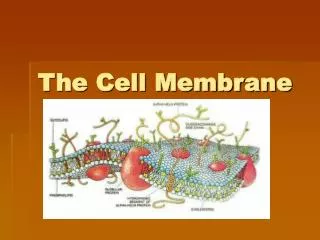
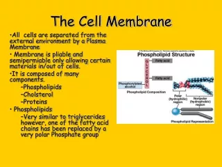
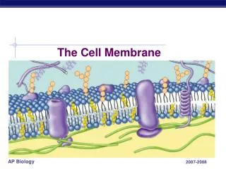
Overview • Cell membrane separates living cell from nonliving surroundings • thin barrier = 8nm thick • Controls traffic in & out of the cell • selectively permeable • allows some substances to cross more easily than others • hydrophobic vs hydrophilic • Made of phospholipids, proteins & other macromolecules
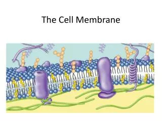
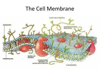
Phospholipids Phosphate • Fatty acid tails • hydrophobic • Phosphate group head • hydrophilic • Arranged as a bilayer Fatty acid Aaaah, one of thosestructure–function examples
Phospholipid bilayer polar hydrophilic heads nonpolar hydrophobic tails polar hydrophilic heads
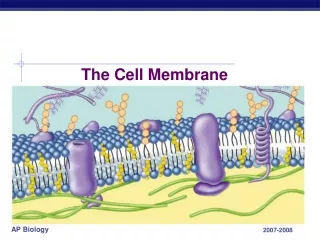
More than lipids… • In 1972, S.J. Singer & G. Nicolson proposed that membrane proteins are inserted into the phospholipid bilayer It’s like a fluid…It’s like a mosaic… It’s the Fluid Mosaic Model!
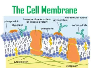
Glycoprotein Glycolipid Transmembrane proteins Peripheral protein Filaments ofcytoskeleton Membrane is a collage of proteins & other molecules embedded in the fluid matrix of the lipid bilayer Extracellular fluid Phospholipids Cholesterol Cytoplasm
Membrane fat composition varies • Fat composition affects flexibility • membrane must be fluid & flexible • about as fluid as thick salad oil • % unsaturated fatty acids in phospholipids • keep membrane less viscous • cold-adapted organisms, like winter wheat • increase % in autumn • cholesterol in membrane
Membrane Proteins • Proteins determine membrane’s specific functions • cell membrane & organelle membranes each have unique collections of proteins • Membrane proteins: • peripheral proteins • loosely bound to surface of membrane • cell surface identity marker (antigens) • integral proteins • penetrate lipid bilayer, usually across whole membrane • transmembrane protein • transport proteins • channels, permeases (pumps)
Why areproteins the perfect molecule to build structures in the cell membrane?
Classes of amino acids What do these amino acids have in common? nonpolar & hydrophobic
Classes of amino acids What do these amino acids have in common? I like thepolar onesthe best! polar & hydrophilic
Proteins domains anchor molecule Polar areas of protein • Within membrane • nonpolar amino acids • hydrophobic • anchors protein into membrane • On outer surfaces of membrane • polar amino acids • hydrophilic • extend into extracellular fluid & into cytosol Nonpolar areas of protein
Porin monomer H+ Retinal chromophore b-pleated sheets NH2 Bacterial outer membrane Nonpolar (hydrophobic) a-helices in the cell membrane COOH Cytoplasm H+ Examples water channel in bacteria proton pump channel in photosynthetic bacteria function through conformational change = shape change
Many Functions of Membrane Proteins Outside Plasma membrane Inside Transporter Enzymeactivity Cell surfacereceptor Cell adhesion Cell surface identity marker Attachment to thecytoskeleton
Membrane carbohydrates • Play a key role in cell-cell recognition • ability of a cell to distinguish one cell from another • antigens • important in organ & tissue development • basis for rejection of foreign cells by immune system
IMMUNOGENETICS • IMMUNITY The immune system in all its form is mankind’s defense mechanism, and in order to understand the inherited disorders of immunity, we must first understand the fundamentals of the genetic basis of immunity. Immune defense mechanism can be divided into two main types: innate immunity – includes a number of non-specific systems which do not require or involve prior contact with the infectious agent. specific acquired or adaptive immunity – involves a tailor-made immune response that occurs after exposure to an infectious agent. Both types can involve either humoral immunity, which combats extracellular infections, or cell-mediated immunity, which fights intracellular infections.
INNATE IMMUNITY • The first simple type of defense against infection is a mechanical barrier. This barrier is represented by skin and by membranes lining the respiratory and gastrointestinal tracts. • If an organism succeeds in invading the body, phagocytosis and bactericidal agents come into effect. HUMORAL INNATE IMMUNITY A number of factors are involved in innate immunity by helping to minimize tissue injury by limiting the spread of infectious microorganisms. These are often called the acute phase proteins and include C-reactive protein, mannose binding protein and serum amyloid P component. In addition, cells when infected by a virus synthesize and secrete interferon , which interferes with viral replication through reducing mRNA stability and interfering with translation.
INNATE IMMUNITY • COMPLEMENT Is a complex series of 20 or so interacting plasma proteins that can be activated by the cell membranes of invading microorganisms, in what is termed the alternative pathway. The various components of complement interact in a specific cascade sequence, resulting in a localized acute inflammatory response through the action of mediators released from mast cells and tissue macrophages. These result in increased vascular permeability and the attraction of phagocytes in the process known as chemotaxis. In addition, the latter components of the complement cascade generate a membrane attack complex that induces defects in the cell membrane, resulting in the lysis of microorganisms. Complement can also be activated through the classic pathway, by the binding of antibody with antigen.
INNATE IMMUNITY • CELL MEDIATED INNATE IMMUNITY PHAGOCYTOSIS Microorganism are engulfed and digested by two major types of cells., either polymorphonuclear neutrophils or macrophages. Polymorphonuclear neutrophils are found mainly in the bloodstream, while macrophages occur primarily in tissue around the basement membrane of blood vessels in connective tissue, lung, liver and in the lining of sinusoids of the spleen and the meddullary sinuses of the lymph nodes. Surface antigen (Ag) on microorganisms result in their being engulfed and fusing with the granules of the phagocytes-which leads to their destruction. EXTRACELLULAR KILLING Virally infected cells can be killed by large granular lymphocytes, known as natural killer cells. Attachment of the natural killer cells to the infected cells results in the release of a number of agents, which in turn results in damage to the membrane of infected cell, leading to cell death.
SPECIFIC ACQUIRED IMMUNITY Many infective microorganisms have, through mutation and selective pressures, developed strategies to overcome or evade the mechanisms associated with innate immunity. There is the need, therefore, to be able to generate specific acquired or adaptive immunity. This can, like innate immunity, be separated into both humoral and cell-mediated processes. HUMORAL SPECIFIC ACQUIRED IMMUNITY The main mediators of this immunity are immunoglobulins or antibodies. Antibodies are able to recognize and bind to antigens of infecting microorganisms. Exposure to a specific antigen results in the clonal proliferation of a small lymphocyte derived from the bone marrow, hence “B” lymphocytes, resulting in mature antibody-producing cells or plasma cells.
HUMORAL SPECIFIC ACQUIRED IMMUNITY • Immunoglobulins The immunoglobulins (Ig), or antibodies, are one of the major classes of serum protein. Their function, both in the recognition of antigenic variability and in effector activities, was initially revealed by protein, and more recently by DNA, studies of their structure. Ig structure – papain (a proteolytic enzyme), splits the Ig molecule into three fragments. Two fragments are similar, each containing an antibody site capable of combining with a specific antigen and therefore referred to as the antigen binding fragment or Fab. The third fragment can be crystallized and was therefore called Fc. The Ig molecule is made up of four polypeptide chains: two ‘light’ (L)-220 amino acid, and two ‘heavy’ (H)-440 amino acid length. They are held together in Y-shape by disulfide bonds and non-covalent interactions. There are five different types of H chain, designated respectively as α,γ,µ,δ, and ε, one each respectively for the five major antibody classes, or what are known as isotypes, IgA, IgG, IgM, IgD and IgE. The L chains are one of two types, either kappa(κ) or lambda (λ), occurring in all five classes of antibody, but with only one type of L chain occurring in each individual antibody. In addition there are four IgG subclasses(IgG1-IgG4), and two IgA subclasses (IgA1,IgA2).
Model of antibody molecule structure. © 2005 Elsevier
Estimated number of the various DNA segments coding for the κ, λ and various heavy chains. Downloaded from: StudentConsult (on 6 October 2006 07:59 AM) © 2005 Elsevier
Classes of human immunoglobulin ------------------------------------------------------------------------------------ Class Mol. Wt. Serum Antibody activity concentration (mg/ml) ------------------------------------------------------------------------------------ IgG 150 000 8 – 16 Binds to microorganisms, neutralizes bacterial toxins IgM 900 000 0.5- 2 Produced in early immune response IgA 160 000 1.4- 4 Guards mucosal surfaces IgD 185 000 0– 0.4 On lymphocyte cell surface, involved in control of activation and suppression IgE 200 000 trace In parasitic and allergic reactions ------------------------------------------------------------------------------------
THE MAJOR HISTOCOMPATIBILITY COMPLEX The major histocompatibility complex (MHC) plays a central role in the immune system. Association of an antigen with the MHC molecule on the surface of the cells is required for recognition of the antigen by the T-cell receptor that, in conjunction with the closely associated protein β2-microglobulin, results in the recruitment of cytotoxic and helper T cells in the immune response. MHC molecules occur in three classes: class I molecules occurring on virtually all cells and which are responsible for recruiting cytotoxic T cells; class II molecules that occur on B cells and macrophages and are involved in signaling T helper cells to recruit further B cells and macrophages; the non-classical class III molecules that include a number of other proteins with a variety of other immunological functions. Structural analysis of the class I and II MHC molecules reveals them to heterodimeres with homology to immunoglobulin. The genes coding for the class I (A,B,C,E,F and G), class II (DR, DQ and DP) and class III MHC molecules, or what is also known as the human leucocyte antigen (HLA)system, are located on chromosome 6.
Transplantation genetics • Replacement of diseased organs by transplantation has become routine in clinical medicine. Except for corneal and boner grafts, the success of such transplants depends on the degree of antigenic similarity between the donor and recipient. Rejection of the transplanted organ or tissue does not occur between identical twins, or between non-identical twins where there has been mixing of the placental circulations before birth. In all other instances, the antigenic similarity of donor and recipient has to be assessed by testing them with suitable antisera or monoclonal antibodies for antigens on donor and recipient tissue. As a general rule, a recipient will reject a graft from any person who has antigens the recipient lacks. • The HLA system is highly polymorphic, two unrelated individuals are therefore very unlikely to have identical HLA phenotypes. The close linkage of the HLA loci means that they tend to be inherited en block, the term haplotype being used to indicate the particular HLA alleles that an individual carries on each of his two number 6 chromosome. • Although crossing over does occur within the HLA region, certain alleles tend to occur together more frequently than would be expected by chance., i.e. they tend to exhibit linkage disequilibrium. An example is the association of the HLA antigens A1 and B8 in populations of Western European origin.
HLA polymorphism and disease associations • A finding which helps to throw light on the pathogenesis of certain diseases is the demonstration of their association with certain HLA types. The best documented is that between ankylosing spondylitis and HLA B27. In the case of narcolepsy, a condition of unknown etiology characterized by a periodic uncontrollable tendency to fall asleep, almost all affected individuals are HLA DR2. • The possession of a particular HLA antigen does not mean that an individual will necessarily develop the associated disease, merely that he or she has a greater relative risk of being affected than the general population. In family, the risks to first-degree relatives of those affected is low, usually no more than 5%. • In general, the mechanisms involved in most HLA disease associations are not well understood.
H-Y antigen • In number of different animal species it was noted that tissue grafts from males were rejected by females of the same inbred strains. These incompatibilities were found to be due to a histocompatibility antigen, known as H-Y antigen. • The H-Y antigen seems, however, to play little part in transplantation in humans. Although the H-Y antigen seems to be important for testicular differentiation and function, its expression does not necessarily correlate with the presence or absence of testicular tissue. • A separate sex-determining region of the Y chromosome (SRY) has been isolated which is now known to be the testis-determining gene.
The Basics Of Blood W.B.C. & Platelet R.B.C. Plasma ANTIBODY Natural & Immune Agglutinins/ Isoantibodies ANTIGEN >400 Agglutinogens on the cell membrane Antigen-Antobody reaction on the cell surface Hemolysis