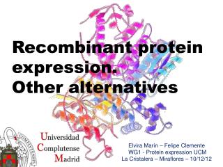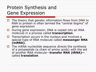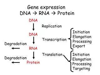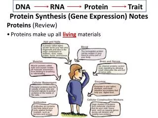
Protein expression
Sources of protein. - purchase from company. - expression systems cloning recombinant proteins into overexpression vector/host systems for intracellular production (bacteria, yeasts, baculovirus, cell free systems, cell cultures). - traditional natural sources . bacterias, animal and plan
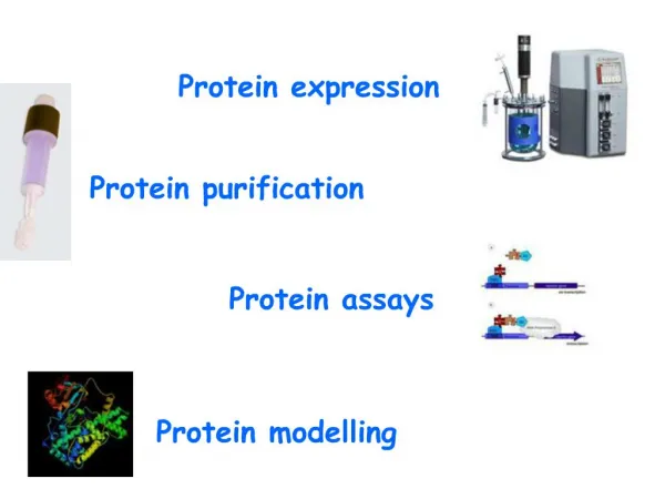

Protein expression
E N D
Presentation Transcript
4. Prokaryotic systems E. coli is a popular and well understood system for heterologous protein expression.
Expression options:
Direct expression. E. coli cytoplasm is a reducing environment - difficult to ensure proper disulphide bonds formation.
Fusion expression.
Ensures good translation initiation. Can overcome insolubility and/or instability problems with small peptides. Has purification advantages based on affinity chromatography.
Secretion
a fusion alternative when proteins are fused to peptides or proteins targetted for secretion. Periplasm offers a more oxidising environment, where proteins tend to fold better. Major drawbacks: limited capacity for secretion (0.1- 0.2% total cell protein compared to 10% produced intracellularly) and inability for posttranslational modifications of proteins.
To minimise proteolysis
For efficient and selective purification
To optimise translation efficiency
5. Disadvantages
6. Expression vector Transcription from the RNA primer promoter, however, is repressed by the product of a trans-acting plasmid gene product, which we have designated rop (for repressor of primer). The rop gene maps downstream from the replication origin in a region that encodes a polypeptide of 63 amino acids whose sequence is completely conserved in pMBI and ColEl. We propose that this polypeptide is the rop gene product and that it regulates plasmid DNA replication by modulating the initiation of transcription of the primer RNA precursor.
Rop-(rop) that encodes an inhibitor of RNA primer transcription. When aplasmid carrying an intact rop gene was present in cells in which /&galactosidase was expressed under the control of the primer promoter, t3-galactosidase- synthesis was reduced to background levels. (cesareni-Proc. NatL Acad. Sci. USA Vol. 79, pp. 6313-6317, October 1982 Genetics)Transcription from the RNA primer promoter, however, is repressed by the product of a trans-acting plasmid gene product, which we have designated rop (for repressor of primer). The rop gene maps downstream from the replication origin in a region that encodes a polypeptide of 63 amino acids whose sequence is completely conserved in pMBI and ColEl. We propose that this polypeptide is the rop gene product and that it regulates plasmid DNA replication by modulating the initiation of transcription of the primer RNA precursor.
Rop-(rop) that encodes an inhibitor of RNA primer transcription. When aplasmid carrying an intact rop gene was present in cells in which /&galactosidase was expressed under the control of the primer promoter, t3-galactosidase- synthesis was reduced to background levels. (cesareni-Proc. NatL Acad. Sci. USA Vol. 79, pp. 6313-6317, October 1982 Genetics)
7. BL21 (DE3) - classical
9. Expression in yeasts
10. Expression in yeasts
13. Baculovirus expression system
14. Advantages X disadvantages
15. Mammalian Cell lines expression systems
17. PromegaPromega
19. No significant differences were observed between untreated plasma (Fig. 1A) and treated samples (Fig. 1B�F). The recovered protein amount in the sample buffer was not measured because of the small volume and the interference of the solubilizing agents. During TCA precipitation, a protein loss probably took place, most likely on account of the acetone-wash step. Thus, the strong
spots representing albumin, heavy and light IgG chains (Fig. 1B) are weaker in comparison with the starting material (Fig. 1A). However, all spots can be detected after easy to perform concentration and desalting step, resulted in a good recovery of all spots (Fig. 1C). Quantitative ammonium sulfate precipitation also resulted in an efficient precipitation of most proteins. The spots representing certain proteins, like complement factor B, C3a and clusterin, are very weak or are missing (the expected position is indicatedin Fig. 1D). The chloroform/methanol method yielded satisfactory results as well, although the spots representing C3a and clusterin are missing in the precipitate (Fig. 1E). Ultrafiltration resulted in recovery of practically all
proteins.
Thus, TCA precipitation, acetone precipitation and ultrafiltration delivered a higher protein recovery compared to ammonium sulfate and chloroform/methanol steps, which also were satisfactory. From the practical point of view, the easiest method to perform is precipitation with TCA, although it usually requires two steps, precipitation with TCA and removal of TCA traces with acetone. Precipitation with acetone requires larger organic solvent volumes (at least threefold of sample volume) and it is inconvenient to perform if the original sample volume is larger than
300l. Ultrafiltration performs very well, however, it is often labor demanding to follow the concentrate volume, the centrifugation steps are usually lengthy, the filters are often
blocked and finally the filtration devices are expensive. Thus, the methods of choice are precipitation with TCA orNo significant differences were observed between untreated plasma (Fig. 1A) and treated samples (Fig. 1B�F). The recovered protein amount in the sample buffer was not measured because of the small volume and the interference of the solubilizing agents. During TCA precipitation, a protein loss probably took place, most likely on account of the acetone-wash step. Thus, the strong
spots representing albumin, heavy and light IgG chains (Fig. 1B) are weaker in comparison with the starting material (Fig. 1A). However, all spots can be detected after easy to perform concentration and desalting step, resulted in a good recovery of all spots (Fig. 1C). Quantitative ammonium sulfate precipitation also resulted in an efficient precipitation of most proteins. The spots representing certain proteins, like complement factor B, C3a and clusterin, are very weak or are missing (the expected position is indicatedin Fig. 1D). The chloroform/methanol method yielded satisfactory results as well, although the spots representing C3a and clusterin are missing in the precipitate (Fig. 1E). Ultrafiltration resulted in recovery of practically all
proteins.
Thus, TCA precipitation, acetone precipitation and ultrafiltration delivered a higher protein recovery compared to ammonium sulfate and chloroform/methanol steps, which also were satisfactory. From the practical point of view, the easiest method to perform is precipitation with TCA, although it usually requires two steps, precipitation with TCA and removal of TCA traces with acetone. Precipitation with acetone requires larger organic solvent volumes (at least threefold of sample volume) and it is inconvenient to perform if the original sample volume is larger than
300l. Ultrafiltration performs very well, however, it is often labor demanding to follow the concentrate volume, the centrifugation steps are usually lengthy, the filters are often
blocked and finally the filtration devices are expensive. Thus, the methods of choice are precipitation with TCA or
21. Eventful Protein Purification
28. Immunoprecipitation
The topic of co-immunoprecipitation (Co-IP) is best preceded by a discussion of immunoprecipitation (IP) to help frame an understanding of the principles involved. Immunoprecipitation is one of the most widely used methods for antigen detection and purification. An important characteristic of IP reactions is their potential to deliver not only the target protein but also other macromolecules that interact with the target.
The principle of an IP is very simple. An antibody (monoclonal or polyclonal) against a specific target antigen is allowed to form an immune complex with that target in a sample, such as a cell lysate. The immune complex is then captured on a solid support to which either Protein A or Protein G has been immobilized (Protein A or G binds to the antibody, which is bound to its antigen). The process of capturing this complex from the solution is referred to as precipitation. Any proteins not �precipitated� by the immobilized Protein A or G support are washed away. Finally, components of the bound immune complex (both antigen and antibody) are eluted from the support and analyzed by SDS-PAGE (gel electrophoresis), often followed by Western blot detection to verify the identity of the antigen.
Traditional immunoprecipitation involves the following steps:
Form the antigen-antibody complex (immune complex) by incubating specific antibody with the antigen-containing sample for 1 hour to several hours.
Capture the immune complex on an immobilized Protein A or Protein G agarose gel support by incubation for 0.5-2 hours.
Remove any non-bound protein (non-immune complex sample components) from the precipitated complex by washing gel support with additional sample buffer.
Boil gel support in reducing SDS-PAGE sample loading buffer.
Recover sample eluted in loading buffer from gel support and analyze by SDS-PAGE.
Perform Western blot analysis, probing with antigen-specific antibody
Co-immunoprecipitation (Co-IP) is a popular technique for protein interaction discovery. Co-IP is conducted in essentially the same manner as an IP. However, in a co-IP�the target antigen precipitated by the antibody �co-precipitates� a binding partner/protein complex from a lysate, i.e., the interacting protein is bound to the target antigen, which becomes bound by the antibody that becomes captured on the Protein A or G beads. The assumption that is usually made when associated proteins are co-precipitated is that these proteins are related to the function of the target antigen at the cellular level. This is only an assumption, however, that is subject to further verification.
Immunoprecipitation
The topic of co-immunoprecipitation (Co-IP) is best preceded by a discussion of immunoprecipitation (IP) to help frame an understanding of the principles involved. Immunoprecipitation is one of the most widely used methods for antigen detection and purification. An important characteristic of IP reactions is their potential to deliver not only the target protein but also other macromolecules that interact with the target.
The principle of an IP is very simple. An antibody (monoclonal or polyclonal) against a specific target antigen is allowed to form an immune complex with that target in a sample, such as a cell lysate. The immune complex is then captured on a solid support to which either Protein A or Protein G has been immobilized (Protein A or G binds to the antibody, which is bound to its antigen). The process of capturing this complex from the solution is referred to as precipitation. Any proteins not �precipitated� by the immobilized Protein A or G support are washed away. Finally, components of the bound immune complex (both antigen and antibody) are eluted from the support and analyzed by SDS-PAGE (gel electrophoresis), often followed by Western blot detection to verify the identity of the antigen.
Traditional immunoprecipitation involves the following steps:
Form the antigen-antibody complex (immune complex) by incubating specific antibody with the antigen-containing sample for 1 hour to several hours.
Capture the immune complex on an immobilized Protein A or Protein G agarose gel support by incubation for 0.5-2 hours.
Remove any non-bound protein (non-immune complex sample components) from the precipitated complex by washing gel support with additional sample buffer.
Boil gel support in reducing SDS-PAGE sample loading buffer.
Recover sample eluted in loading buffer from gel support and analyze by SDS-PAGE.
Perform Western blot analysis, probing with antigen-specific antibody
Co-immunoprecipitation (Co-IP) is a popular technique for protein interaction discovery. Co-IP is conducted in essentially the same manner as an IP. However, in a co-IP�the target antigen precipitated by the antibody �co-precipitates� a binding partner/protein complex from a lysate, i.e., the interacting protein is bound to the target antigen, which becomes bound by the antibody that becomes captured on the Protein A or G beads. The assumption that is usually made when associated proteins are co-precipitated is that these proteins are related to the function of the target antigen at the cellular level. This is only an assumption, however, that is subject to further verification.
32. Bud server kterej uz ma matrice predchozich struktur � swissprot a PDBviewerBud server kterej uz ma matrice predchozich struktur � swissprot a PDBviewer
33. The complete set of proteins expressed and modified by a cell, tissue or organism at a given time.
Proteins are the physical interface between genome and environment.
35. Enormous dynamic range (1 to 108)
Wide range of molecular weight (750 kDa to 350 Da)
Of pI (3 to 12)
Of hydrophobicity
Various stabilities