Download
1 / 46
460 likes | 614 Vues
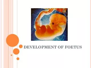
DEVELOPMENT OF FOETUS. INTRODUCTION. Life begins at the time fertilization of the ovum The zygote formed is a single cell which develops into fully formed adult Prenatal development is the process in which an embryo or fetus gestates during pregnancy, from fertilization until birth.

E N D
INTRODUCTION • Life begins at the time fertilization of the ovum • The zygote formed is a single cell which develops into fully formed adult • Prenatal development is the process in which an embryo or fetus gestates during pregnancy, from fertilization until birth.
Gestation Period • The gestation period of • Germinal Period - This begins at fertilization and extend till the third week. • Embryonic period – This extend from 4th ~ 8th week, involving changes in shape and external appearance. • Fetal period – This extends from 3rd month upto termination of pregnancy.
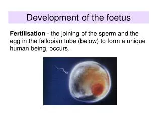
Development of supportive structures of fetus & Major Events • Fertilization • Clevage Division – The process of repeated mitotic division results in increase of number of cells known as Clevage give rise to blastomere. • Formation of morula and blastocyst - blastomere form compact ball called morula. The centre of morula is inner cell mass. Blastocyst – The cells of morula continue to divide and forms blastocele. At this stage zygote is blastocyst. Outer cell is trophoblast and inner cell mass is embryoblast. • Implantation of blastocyst – Blastocyst attaches to endometrium and implantation takes place on 6th ~ 7th day after fertilization. Now Endometrium is ready to support the pregnancy, and is now referred as the decidua .
The Decidua • The corpus luteum continues to produce progesterone which stimulates secretery activity of endometrial glands and increase the size of blood vessel and form soft spongy bed. The layers of Decidua • Basal Layer – Lies above the biometrium. It contains basal portion of glands • Functional Layer – It is through this layer that the cleavage of placental separation occur • Superficial compact layer – This forms surface of decidua. After implantation of the blastocyst the decidua is renamed as • Basal decidua – The decidua underneath the blastocyst. • Caspular decidua – It thin superficial layer covering the ovum. • Parietal decidua – the rest of decidua lining the uterine cavity outside the site of implantation. Functions • Provides bed for implantation • Supplies nutrition to early stage of growing ovum • Protective function
Formation of germ layer • This is first major event after implantation. • Three layers are formed – Ectoderm – layer forms skin and nervous system. Mesoderm – Layer forms bone, muscle, heart and blood vessel. Endoderm – Forms mucus membrane and glands • The three layer together are known as embryonic plates.
Development of Placenta • Placenta develops from two sources Fetal part from chorionfrondosum and maternal part from decidua basalis. STAGES • Implantation • Changes to decisdua • Trophoblast forms chorion • Chorionic villi are formed i.e. finger like projection • Villi becomes profused in the basal decidua and eventually develops into placenta • Villi under capsular decidua being less nourished degenerates and forms chorionic laeve which is origin of chorionic membrane • Chorionic villus centre consist of mesoderm and fetal blood vessel and branches of umbilical arteries and vein.
Placental Circulation Maternal Placental circulation • The maternal blood through spiral arteries comes to placental bed and surrounds the inter villus space and from there enters the veins and is drained by them. Fetal Placental Circulation • Fetal blood comes to placenta through umbilical Arteries and enters the chorionic villi. • Veins from chorionic villi drain into umbilical vein which carries the blood from fetus to placenta.
Functions • Respiration – Oxygen is obtained and carbon dioxide is excreted through placenta. • Nutrition - Placenta provides proteins as amino acids, carbohydrates and fats as fatty acids. • Storage – It stores glucose as glycogen. • Excretion – It gives out carbon dioxide bilirubin urea uric acid • Protection – Prevents entry of micro organism and noxious agent but not drugs and virus. • Endocrine – Produces human Chorionic Gonadotrophin, Estrogen & Progestron
Anatomic Variations • Succenturate Lobe of placenta , Small extra lobe is present. • Circumvallate placenta – in this an opaque ring is seen on fetus surface. • Battle dore insertion of cord – in this umbilical cord is attached closed to margin of placenta • Velamentous insertion of chord – Chord is inserted into membrane some distance away from edge of placenta. • Placenta preavia - Implantation in the lower part of uterine cavity. • Bipartite Placenta – to complete and separate lobes are present • Tripartrite Placenta – Three lobes are present
Umbilical Cord • The umbilical cord or funis forms the connecting link between the fetus and placenta. • It has two arteries and one vein. • It is protected by whartsons jelly average length 50 cm • If cord is long may become wrapped around the neck or body of fetus
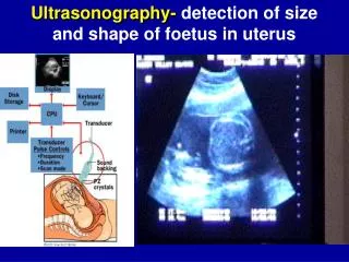
Amniotic Fluid • The source is through both fetal and maternal • It is secreted by amnion • Some fluid is oozed from maternal vessel and some from fetal vessel • Fetal urine also contributes after 10th week of gestation • It is clear, pale, straw colored fluid consisting 99% of water.
Functions • Provides protection for embryo against shock, blow or pressure. • Embryo floats in the fluid. • It equalizes pressure • Allows fetal movement • Maintains constant temperature • Aids in effacement of cervix and dilation of uterine os. • Provides small amount of nutrient
The Fetal Membrane • Has two layers Chorion – It is thick opaque membrane derived from trophoblast. Amnion – It is inner layer, It is smooth, tough, translucent membrane derived from inner cell mass.
Changes by weeks of gestational age Germinal period (0~3 Weeks) 1st week - Clevage division - formation of morula & blastocyst - Implantation of Blastocyst 2nd week - Implantation is complete - formation of biliminar embryonic disc. - Amniotic cavity - Amnion - Yolk sac - Connecting stalk - Chorionic sac
3rd week - formation of trilaminar germ disc - Primitive streak - Notochord Gastrulation This is a process by which bilaminar germ disc is converted into trilaminar embryonic disc. Primitive streak appears at the 15th day. It is narrow median groove formed by ectodermal cell. A neural groove ( future spinal cord forms over the notochord with brain bulge at one end) Somites, the division of future vertebra form. It is formed by proliferation of mesoderm into paired cuboidal bodies called somites. Primitive heart tube is forming. Vasculature begins to develop
Embryonic Period (4th -8th week)week -4 • Three Germ layer appears • Folding of the embryo(Begins to curve into C- shape) • Heart further develops • Brachial arches, grooves will form • Neural tube closes • Ear begins to form as otic pit • Arm buds and tails are visible • Pulmonary primordium appears • Hepatic plate appears • Cystic diverticulum, which will become gall bladder and pancreatic bud will appear • Urorectal septum begins to form • Anterior and posterior horn differentiate into spinal cord • Spleen appears • Uretric buds appears
Week-5 • Embryo measures 8 mm in length • Lens pits and optic cups form • Nasal pit forms • The brain develops • First heart beat begins and four chambers are functioning • Leg buds form and hands form as flat paddles on the arms • Umblical cord develops
Week-6 • Embryo measures 13-mm • Lungs begins to form • Arms and legs have lengthened • Hands and feet have digits • Gonadal ridge begins to perceptible • Lymphatic system begins to develop • Nostrils forms • Intestine and pancreas grow
Week-7 • Embryo measures 18 mm • Nipples & hair follicles begins to form • Location of elbow & toes are visible • Teeth begins to develop • All essential organ have atleast begun formation
Week-8 • Cartilage & bones begins to form • The tongue begins to develop • Intestine moves out of the umbilical cord in to the abdomen • Facial features continues to develop • Sex differentiation begins • Embryo measures 30 mm and weight is 1 gm
Fetal period • The fetal period is extending from the beginning of the third month(9th week) up to the termination of pregnancy • Associated with complete development of placenta, umbilical cord & fetal membranes • Developing organism is called fetus
Week-9 • Fetus is about 50 mm long and weight is 8 gms • Head constitutes half the crown heel length of fetus • Face is broad, eyes get widely separated, ears are low set and eye lids gets fused • Movement begins • Most of the joints are formed
Week 10 • Face has human profile • Placenta begins to function • Genitalia have male or female characterstics but still not fully formed Week 11 • Genitals appears well differentiated • Hair and nails begins to grow • Fingers and toes are separated • Amniotic fluid begins to accumulate as the kidneys begins to function
Week-12 • Length is about 8 cm and weight is 25 gms • Eyes are widely spaced • Vocal cords begins to form • Heart beat is audible by droppler • Pancreas are active • RBCs are produced in liver Week-13 • Growth is rapid • Inhaling and exhaling have started • Neck is getting longer and hands becoming more functional
Week-14 • Fetal skin is almost transparent • More muscle tissues and bones have developed • Thyroid gland has matured and fetus begins to produce hormones in boys prostate gland and in females the ovaries move from abdomen to pelvis • Fine hair called lanugo develops
Week-15-16 • Sucking motions are made by mouth • Fetus makes active movements • Tiny bones in the middle ear begins to harden • Ovaries are differentiated and contains primary ovarian follicles • Meconium is made in intestinal tract • Liver and pancreas are functioning • By end of 16th week sucking, swallowing and blinking are evident
Week 17-20 • Lanugo covers the entire body • Eyebrows & eyelashes appear • Mother feels fetal movements, known as quickening • Nails appears on fingers & toes • Skin is covered with vernix caseosa • Brown fat is formed & is site of heat production • By 18th week, uterus is formed & canalization has begun • By 20th week testes begins to descend • Fetal heart beat can be heard with stethoscope • Immunities are transferred from mother to fetus
Week 21-25 21 • Shows substantial gain in weight • Rapid eye movement begins • WBCs are under production • More amniotic fluid is swallowed 22 • Eye lids & eye brows are fully formed • Liver breaks down bilirubin 23 • Proportion of body are quite similar to a new born • Fetus is able to hear
24 • Baby is officially considered viable • By end of this week fetus has grown 28-36 cm & weighs 550 gms • Pupils reacts to light • Taste buds begins to form • Production of lung surfactant begins 25 • Structure of spine begins to form the joints, ligaments and rings • Blood vessels of lungs develop • Nostrils begins to open
Week 26-29 26 • Fetus touches to a length of about 35-38cm & weighs about 1200gms • Brain develops rapidly • Retina begins to form • Air sacs in lungs forms now • Eye lids open in 26th week 27 • Response to sound grows • The central nervous system matures • Lungs continue to grow & is ready to function outside of the womb • Retina have formed
28 • Eyelids open & close • Fetal body is getting plump & rounded • Muscle tone is improving 29 • Fetal head is in proportion with body now • Fat accumulate under the skin • Movement is increased • Fetal spleen is important site of haemtopoesis • Male testes descend in scortum & female clitoris is prominent.
Week 30-34 • Length is about 38-43 cm weight is about 1,600 gms • As fetus continues to grow the amniotic fluid will decrease • Early lanugo disappears slowly & own hair may begin to appear • Bone marrow produces RBCs 31 • Physical growth slows down • Fetus gains weight • Bones are fully developed, but are still soft and pliable .
32 • Movements are bit reduced because of increased size • All five senses are working as thalamic brain connections which mediates sensory input form 33 • Active mores reflex is present • The skull is quite pliable & not completely joined 34 • In male fetus the testicles descend into the scortum • Eyes are opened wide when awake & closed while sleeping • Fetus has already turned to a head down position
Week 35-40 • Fetus reaches a length of about 40-46 cm & weight is around 1,800-1,900 gms • Lanugo disappear • Fetus takes up the space in most of uterus & mother may feel like chut has run out of room 36 • Fetus may descend into the birth canal called lightening • Skin is smooth & pink • Fully developed pair of kidneys
37 • Fetus is official full term • Length reaches up to 42-48 cm & may weigh 1,800-2,700 gms • Grasp reflex is firmly established 38 • Birth position is usually assumed • Fetal intestines are accumulating lots meconium • Hairs are thick • Circumference of head & abdomen are about same size
39 • Lanugo has gone except on the upper arm & shoulders • Lungs are matured and surfactant production is increased • Fat stores are more that will help to regulate the body temperature after birth • Fetus is around 50 cm in length & weighs about 3288 gms
Week 40 • Much of vernix have disappeared • 15 % of baby's body is fat • Breast buds are present. • Lungs will continue to develop until birth • Fetus is 51cm &weight is around 3400 grams. • Any time & any day the fetus is delivered now and the development continues postnatal with child development stages
CONCLUSION • The development of fetus is wonderfully &precisely coordinated series of events. Human development is an ongoing process of transformation that begins with fertilization & continues through teenage years and beyond .