Download
1 / 103
1.13k likes | 1.55k Vues
Case 1: Diagnosis and Management of Head Trauma. Lee Chuy, Katherine Lee, Sidney Albert. Demographic Data. GT 22 year old Male Side swept by 10 wheeler truck while walking along Forbes St. and taken by bystanders to USTH. PE. Vital Signs BP: 110/70 PR: 95/min RR: 20/min. PE. Skin

E N D
Case 1: Diagnosis and Management of Head Trauma Lee Chuy, Katherine Lee, Sidney Albert
Demographic Data • GT • 22 year old • Male • Side swept by 10 wheeler truck while walking along Forbes St. and taken by bystanders to USTH
PE • Vital Signs • BP: 110/70 • PR: 95/min • RR: 20/min
PE • Skin • 5 cm laceration on left temporal area • Multiple abrasions and lacerations over the face and extremities
PE • Neurologic • Confused • Spontaneous eye opening • Localizes upon pain stimuli • Pupils 2mm, ERTL
Imaging • X-ray • Linear fracture over left temporal area
After X-ray • Less responsive • Left extremity withdraws to pain • Right in extension • Eye opening on painful stimuli • No verbal output • Pupils: 3mm, RTL, right; 5mm, non-reactive, left
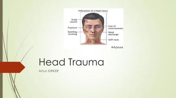
Head Injury • A head injury is any trauma that leads to injury of the scalp, skull, or brain • The injuries can range from a minor bump on the skull to serious brain injury. http://www.nlm.nih.gov/medlineplus/ency/article/000028.htm
Closed head injury • hard blow to the head by striking object, but object did not break the skull • Open or Penetrating head injury • hit with an object that broke the skull and entered the brain • which the dura mater, the outer layer of the meninges, is breached • Compressive head injuries • The Skull is compressed between two forces. Massive injury that usually results in instantaneous death http://web.archive.org/web/20030821142023/http://cats.med.uvm.edu/cats_teachingmod/pathology/path302/np/home/neuroindex.html
Primary injury is induced by mechanical force and occurs at the moment of injury. • Secondary injury is not mechanically induced. It may be delayed from the moment of impact, and it may superimpose injury on a brain already affected by a mechanical injury
Primary injury • 2 main mechanisms that cause primary injury • contact (eg, an object striking the head or the brain striking the inside of the skull) , may result in injury to the scalp, fracture to the skull, and surface contusions • acceleration-deceleration, results from unrestricted movement of the head and leads to shear, tensile, and compressive strains. These forces can cause intracranial hematoma, diffuse vascular injury, and injury to cranial nerves and the pituitary stalk. • Contusions are distinct areas of swollen brain tissue.They are typically found on the poles of the frontal lobes, the inferior aspects of the frontal lobes, the cortex above and below the operculum of the sylvian fissures, and the lateral and inferior aspects of the temporal lobes.
Secondary injury • Secondary injury may occur hours or even days after the inciting traumatic event. • Injury may result from impairment or local declines in cerebral blood flow (CBF) after a TBI. • Decreases in CBF are the result of local edema, hemorrhage, or increased intracranial pressure (ICP). As a result of inadequate perfusion, cellular ion pumps may fail, causing a cascade involving intracellular calcium and sodium. Resultant calcium and sodium overload may contribute to cellular destruction. • Excessive release of excitatory amino acids, such as glutamate and aspartate, exacerbates failure of the ion pumps. • As the cascade continues, cells die, causing free radical formation, proteolysis, and lipid peroxidation. • These factors can ultimately cause neuronal death.
The exact role of the inflammatory response in secondary injury is not known. However, it is believed to contribute to cell damage.9 • Clinical conditions associated with the risk of a decreased CBF are arterial hypotension, hypoxemia, intracranial hemorrhage and malignant brain edema, and hyperthermia.
Focal or Diffuse • These 2 forms of injury are commonly found together. • Focal injuries include scalp injury, skull fracture, and surface contusions and are generally be caused by contact. • Diffuse injuries include DAI, hypoxic-ischemic damage, meningitis, and vascular injury and are usually caused by acceleration-deceleration forces.
http://emedicine.medscape.com/article/326643-overview • Classification and Complications of Traumatic Brain Injury • Author: Percival H PangilinanJr, MD • April 2, 2008
2. Differentiate primary brain injury from secondary brain injury.
Primary Brain Injury • physiological and anatomic damage to the brain, occurring at the superficial layers of the brain and extending inward as mechanical force increases. • Results from the initial trauma to the brain
Primary Brain Injury • Intracerebral hemorrhage • Subdural hemorrhage • Subarachnoid hemorrhage • Epidural hemorrhage • Cerebral contusion • Cerebral laceration • Axonal stretch injury
Primary Brain Injury • classified as penetrating or closed, depending on the mechanism of injury: 1. Penetrating injuries are characterized by the velocity of the missile at impact and the location of the missile impact: • The higher the velocity of the missile, the more severe the injury. • The most severe injuries are caused by high-velocity bullets • The lower the missile tract, such as in the brainstem, basal ganglia, or large artery, the more severe the injury.
Primary Brain Injury 2. Closed injuries are classified as mild, moderate, or severe, depending on the neurological status of the patient soon after the injury . -Moderate or Severe Closed Injuries are characterized by the velocity and vector of forces applied to the head, such as motor vehicle accidents, assaults, or falls. - There are two types of closed injuries. a. "Coup" injuries, caused by the impact of the head with an obstacle b. "Contrecoup" injuries, caused by the brain rebounding within the skull from the initial "coup" injury.
Secondary Brain Injury • Subsequent neuronal damage due to the sequelae of trauma Mechanisms of secondary injury • hypoxia • hypotension • hydrocephalus • intracranial hypertension • intracranial hematoma Schwartz’s Principles of Surgery, 9th ed
The focus of basic research into brain trauma, critical care medicine, and neurosurgical intervention is to decrease the effects of secondary injury Schwartz’s Principles of Surgery, 9th ed
ABCDs of resuscitation • Airway • Breathing Assessed and stabilized • Circulation • Hypoxia and hypotension worsen outcome in traumatic brain injury due to secondary injury making cardiopulmonary stabilization critical • Disability (GCS score) Schwartz’s Principles of Surgery, 9th ed
Glasgow Coma Scale or GCS is a neurological scale that aims to give a reliable, objective way of recording the conscious state of a person for initial as well as subsequent assessment.
GCS was initially used to assess level of consciousness after head injury, and the scale is now used by first aid, EMS, and doctors as being applicable to all acute medical and trauma patients. In hospitals it is also used in monitoring chronic patients in intensive care.GCS is used as part of several ICU scoring systems, including APACHE II, SAPS II, and SOFA, to assess the status of the central nervous system.
Generally, brain injury is classified as: • Severe, with GCS ≤ 8 • Moderate, GCS 9 - 12 • Minor, GCS ≥ 13.
Patients score • EYE • Spontaneous eye opening • Score of 4 • VERBAL • Noted to be confused • Score of 4 • MOTOR • Localizes on painful stimuli • Score of 5 • GCS total of 13 • BRAIN INJURY
On the way back… • EYE • Eye opening on painful stimuli • Score of 2 • VERBAL • No verbal output • Score of 1 • MOTOR • Withdraws to pain • Score of 4 • GCS total of 7 • SEVERE BRAIN INJURY
Brain herniation syndromes Lim, John Harold Lim, Mary
Brain Herniation • Brain tissue, cerebrospinal fluid, and blood vessels are moved or pressed away from their usual position in the head. • Occurs when something inside the skull produces pressure that moves brain tissues. • Most often the result of brain swelling from a head injury.
General symptoms indicating brain herniation • Cardiorespiratory distress • Coma • Irregular breathing • Irregular pulse • Loss of all brainstem reflexes (blinking, gagging, pupils reacting to light) • Progressive loss of consciousness
5 main types of brain herniation syndromes • TRANSTENTORIAL HERNIATION • ALAR OR SPHENOID HERNIATION • SUBFALCINE HERNIATION • FORAMEN MAGNUM HERNIATION • EXTRACRANIAL (Calvarial) HERNIATION
Herniation 1. &2. Descending transtentorial herniation 3. Subfalcine Herniation 4. Extracranial (calvarial) herniation 5. Ascending transtentorial (upward) herniation 6. Foramen magnum (tonsillar) herniation
Herniation • Alar (Sphenoid) herniation
Descending transtentorial herniation • Caused by mass effect in the cerebrum which pushes the supratentorial brain through the incisura to the posterior fossa • Clinical presentation includes occulomotor (CN III) paresis, Mydriasis (ipsilateral dilated pupil), abnormal EOM's, contralateral hemiparesis, and at times ipsilateral hemiparesis
Descending transtentorial herniation • Imaging findings of descending transtentorial herniations include ipsilateral ambient cistern widening and ipsilateral prepontine cistern widening. A contralateral temporal horn is also widened.
Descending transtentorial herniation • ipsilateral ambient cistern widening
Descending transtentorial herniation • ipsilateral prepontine cistern widening
Descending transtentorial herniation • Widened contralateral temporal horn
Ascending transtentorial herniation • Less common then descending transtentorial herniation. • Usually caused by a slowly growing cerebellar or brainstem process, such as a diffusely infiltrating astrocytoma. • Clinical presentation includes nausea and vomiting followed by obtundation
Ascending transtentorial herniation • “Spinning top” appearance of the midbrain • Due to compression bilaterally on the posterolateral aspects of the midbrain as the posterior fossa squeezes through the incisura from below.
Ascending transtentorial herniation • Subsequent narrowing of the bilateral ambient cisterns as the cerebellar tissue extends through the ambient cisterns. • Finally, there is filling of the quadrigeminal plate cistern as the cerebellum again persistently extends upward through the incisura.
Alar or Sphenoid herniation • Clinical symptoms are generally minimal. • Associated with other herniations such as subfalcine or transtentorial herniations which are more clinically apparent. • Divided into posterior and anterior herniations. • Frontal lobe masses will cause a posterior alar herniation as the frontal lobe extends posterior and inferiorly over the sphenoid ridge. • Temporal lobe lesions or lesions of the insula can cause anterior alar herniations. This occurs as the temporal lobe extends superiorly and anteriorly over the sphenoid bone.
Alar or Sphenoid herniation • Imaging findings of alar herniation are best demonstrated utilizing the middle cerebral artery. • anterior or posterior displacement of the middle cerebral artery • CT: Anterior left alar herniation • Cerebellum filling the quadrigeminal plate cistern and ambient cisterns from an ascending transtentorial herniation
Subfalcine herniation • Occur as the brain extends under the falx in the supratentorial cerebrum. • Occur in conjunction with transtentorial herniation or in isolation. • Can present clinically as headache. Later on as the herniation progresses, contralateral leg weakness can occur.