
WT R6/2
7Q 109Q. Human brain ( frontal cortex ; anti-p-AMPK ) . A. Non-HD. HD patient. - + - + AICAR ( 1mM) . C. D. AMPK α -p. AMPK α 1. Actin. E. F.

WT R6/2
E N D
Presentation Transcript
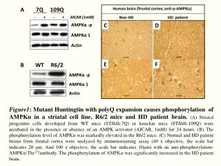
7Q 109Q Human brain (frontal cortex; anti-p-AMPK) A Non-HD HD patient - + - + AICAR (1mM) C D AMPKα -p AMPKα 1 Actin E F Figure1: Mutant Huntingtin with polyQ expansion causes phosphorylation of AMPKα in a striatal cell line, R6/2 mice and HD patient brain.(A) Striatal progenitor cells developed from WT mice (STHdh-7Q) or knockin mice (STHdh-109Q) were incubated in the presence or absence of an AMPK activator (AICAR, 1mM) for 24 hours. (B) The phosphorylation level of AMPKα was markedly elevated in the R6/2 mice. (C) Normal and HD patient brains from frontal cortex were analyzed by immunostaining assay (40 x objective, the scale bar indicates 20 μm. And 100 x objective, the scale bar indicates 10μm) with an anti-phosphorylation-AMPKα-Thr172antibody. The phosphorylation of AMPKα was significantly increased in the HD patient brain. B WT R6/2 AMPKα -p AMPKα 1 Actin
A B 7Q 109Q * 250 200 150 DCFDA Fluorescence Intensity 100 50 0 109Q 7Q 300 * STHdh-109Q * 250 250 200 a 200 Compound C (10 mM)- + - b NAC (5 mM)- - + 150 a 150 b C H2O2 (mM) 0 10 100 500 D DCFDA Fluorescence Intensity AMPKα-p DCFDA Fluirescence Intensity 100 100 AMPKα -p AMPKα 1 50 AMPKα 1 50 Actin Actin 0 0 H2O2 (100 μM)- + + + pcDNA3.1 WT 172T/D 172T/A E NAC (5mM)- - + - AMPKα1 Compound C (10 μM)- - - + Figure 2: Mutant Htt-expressing cells contain higher levels of ROS and accumulate more ROS upon oxidative stress in AMPK dependent manner, and elevation of ROS caused the phosphorylation of AMPKα in the presence of mutant Htt. Striatal knock-in cells were used to study this free radical production. Measurement of ROS level was conducted by incubating the indicated cells with DCF-DA for 30 min. The relative levels of DCF fluorescence were quantified using a fluorescence reader. (A) The mutant Htt-expressing cells contain higher levels of ROS. (B) Expression of AMPK-Thr172D dramatically increased ROS in the presence of mutant Htt express cells, and free radical production was positively correlated with AMPK activation in the presence of mutant Htt. (C) STHdh-7Q and STHdh-109Q cells were pre-treatmented with NAC and compound C for 1h, followed by a 24-hr treatment with H2O2 (100 M). NAC (N-acetylcysteine) is an antioxidant, and compound C is an AMPK inhibitor. The mutant Htt expressing cells were accumulated more ROS upon oxidative stress, and this phenomenon can be reduced by NAC and compound C. (D) STHdh-109Q cells were treated with H2O2 and incubated for 24h, the phosphorylationof AMPKα were significantly increased. This suggests that the activation of ROS subsequently promotes the activation of AMPKα. (E) STHdh-109Q cells were treated with NAC and compound Ctheover-activation of AMPKα was dramatically reduced.
7Q 109Q 7Q+CC 109Q+CC 25Q 109Q A B 120 STHdh cells 7Q 109Q 100 80 * MTT Assay (% control) 60 40 20 0 control AICAR 5mM Flow Cytometric Assay C D Flow Cytometric Assay 60 ST14A cells * 120 H2O2 50 100 * 40 80 * 30 Cell death (%) Control H2O2 AICAR+H2O2 20 60 MTT Assay (% control) 10 40 0 20 * * * 0.001 0.01 0.1 1 10 100 0 AICAR (mM) Figure 3: Activation of AMPK cause higher cell death in the presence of mutant Htt . AMPK inhibitor protects striatal cells from death caused by free radical production.Cell survival was determined using the MTT and Flow Cytometric assay. Cell viability is shown as the percentage observed in the control groups. (A) ST14A cells were transfected with the indicated poly-Q-construct and incubated with AICAR for 24h. (B) STHdh-7Q and STHdh-109Q cells were treated with AICAR for 24h. Both in the figure A and figure B, activation of AMPK significantly increased cell death in the presence of mutant Htt. (C) Striatal progenitor cells were pretreated with AICAR or H2O2 for 24hr were significantly increased cell death, and cells were co-treat with AICAR and H2O2 was further enhanced cell death. (D) Striatal progenitor cells treated with or without compound C for 1 hr, followed by a 24 hr treatment with H2O2 (100 M). The mHtt expression cells more sensitive to oxidative stress and Compound C can protect striatal cells from death.
Wild-type R6/2 mice mHtt AMPKa-p mHtt AMPKa-p Hoechst Merge Hoechst Merge A B R6/2 (weeks) 7 10.5 12 AMPK-pT172 C AMPKα1 Control AICAR 1mM STHdh7Q/7Q STHdh7Q/7Q7 Lamin A/C AMPKα-p Hoechst Merge AMPKα-p Hoechst Merge STHdh109Q/109Q STHdh109Q/109Q AMPKα-p Hoechst Merge AMPKα-p Hoechst Merge D Control AICAR 1mM STHdh7Q/7Q STHdh7Q/7Q AMPKα1 Hoechst Merge AMPKα1 Hoechst Merge STHdh109Q/109Q STHdh109Q/109Q AMPKα1 Hoechst Merge AMPKα1 Hoechst Merge Figure 4: PhosphorylatedAMPKα was enriched in the nucleus in a stritum cell line and in the striatum of 12-weeks old R6/2 mice. (A) The phosphorylatedAMPKα were expressed in the cytoplasm in WT mice, but not in the HD mice. On the contrary, phosphorylatedAMPKα was enriched in the nucleus of R6/2 mice. Moreover, the phosphorylated AMPK co-localize with mutant Htt in the nucleus. (B) the nuclear enrichment of phosphorylatedAMPKαis stage dependent. (C,D) The phosphorylatedAMPKα were expressed in the cytoplasm in 7Q cells. But the majority of phospho-AMPKαare restricted in the nucleus in 109Q cells. AICAR treatment led to the nuclear enrichment of activated AMPKα. Scale bar indicates 20μm.
Figure 5: Abnormal nuclear accumulation of AMPKα in brains of HD patients.Significant amounts of AMPKα were found in nucleus of HD patients, but not in the age-matched controls. Scale bars indicate 20 μm. A. Caudate nucleus B. SubstantiaNigra Non-HD-2 AMPKα1 Hoechst Merge HD patient-1 AMPKα1 Hoechst Merge C. SubstantiaNigra Non-HD-5 AMPKα1 Hoechst Merge Non-HD-6 AMPKα-p Hoechst Merge HD patient-3 HD patient-4 AMPKα1 Hoechst Merge AMPKα-p Hoechst Merge
Transfection: AMPKα1-T172D-NES/V5 A B Post-nucleus nucleus STHdh-7Q AICAR (mM) 0 1 1 1 0 1 1 1 Time (h) 24 6 12 24 24 6 12 24 Hoechst Merge Anti-V5 AMPKα -p AMPKα 1 STHdh-109Q STHdh-109Q β -tubulin Hoechst Merge Anti-V5 120 109Q Lamin A/C Transfection: AMPKα1-T172D-NLS/V5 109Q+AICAT 100 AMPKα -p STHdh-7Q 80 STHdh-7Q AMPKα 1 60 MTT Assay (%) Hoechst Merge Anti-V5 40 STHdh-109Q 20 C D Hoechst Merge Anti-V5 0 WT 172T/D 172T/D-NES AMPKα 1 172T/D 172T/D-NLS 172T/D-NES AMPKα 1 Figure 6: Nuclear enrichment of AMPKα is activity-dependent and the nucleus translocation is important to the toxic effect of AMPK.(A) Large amount of the phosphorylatedAMPKα can be found in the nuclear fraction of the 109Q cells, but not in the naïve 7Q cells. (B) The NES domain in AMPKα1-Thr172 D is responsible for the cytoplasmic retention of V5-positive signal and a NLS domain substitution can tranlocatethe V5-positive signal to the nucleus. (C, D) cell survival rate was significantly increased with AMPKα1-Thr172 D-NES compared to those with the AMPKα1-Thr172 D-NLS, but when we treat cell with AICAR the MTT reader were reduced.
A B WT HD 7Q 109Q C N C N Compound C - + - - - + - - P300S89 NAC - - 5 1 - - 5 1 β -tubulin P300S89 Actin Lamin A/C C D Non-HD-5 n=~250/patients P300-s89 Hoechst Merge HD patient-3 P300-s89 Hoechst Merge Figure 7: Mutant Htt enhances the AMPK-mediate phosphorylation of p300 at serine89 in a striatal cell line, the striatum of R6/2 and the human brain.(A) the phosphorylation level of nuclear p300 was markedly elevated in R6/2 mice compared to wild-type ones. (B) mHttexpressing cell showed higher P300-S89 phosphorylation which is decreased by the treatment of AMPK inhibitor and antioxidant.(C) Large amounts of P300-S89 were found in nucleus of HD patients, but not in the normal human. Scale bars indicate 20 μm. (D) Results of quantitation, the P300-S89 phosphorylation profile in nucleus were increased in HD patients.
A B * 7Q 109Q * - CC A - CC A C Bcl2 Actin Figure 8: Mutant Htt enhances the AMPK-mediate Bcl2 gene expression in a striatal cell line the striatum of R6/2 mice. (A, B) Striatal progenitor cells and WT/HD mice were harvested for total RNA collection and used for the quantitative RT-PCR analysis. Expression levels of Bcl2 were normalized to that of GAPDH. The mutant Htt-expressing cells and R6/2 mice possess lower levels of Bcl2 mRNA expression. Data are expressed as the mean ± SEM of three independent experiments. (C) STHdh-7Q and 109Q cells were treated with compound C or AICAR and incubated for 24h, mHtt expressing cell showed lower Bcl2 which could be enhanced by treating with AMPK inhibitor.
A B F Control 120 7Q 109Q 100 AMPK α -p Hoechst Merge 80 AICAR b a MTT Assay (% control) 109Q 7Q 109Q 60 C D - - - FK + - + - AICAR (1mM, 24h) Q7 CON CGS H89 KT KT AMPK α -p Hoechst Merge 40 24h 6h 1h - - - - - CGS (10μM) - - - 10 1 10 PKA inhibitor (μM) CGS 20 AMPKαT172 AMPK αT172 0 CGS - + - + + + AMPK α -p Hoechst Merge 1.2 2.0 1.7 1.4 3.7 3.1 3.7 1 (AMPK-P/total) 1 2.25 1.5 2.37 2.07 2.42 (AMPK-P/total) AICAR - - + + + + H89 - - - - 1 10 AMPKα 1 AMPK α 1 Actin Actin E F Figure 9.Stimulation of the A2A adenosine receptor suppresses AMPK activation and rescues neuronal damage in a PKA-dependent pathway. (A) The mHttexpressing cells are more sensitive to cell damage induced by AMPK and when cells pretreated with CGS were rescues neuronal death. (B) The activation of AMPK significantly increased cell death whichis mildly rescued by CGS. PKA inhibitor reduced the CGS protective effect. (C) mHttexpressing cells showed higher AMPKα-T172 phosphorylation which is decreased by CGS and FK. PKA inhibitors including H89 and KT rescuethis phenomenon (D). (E)109Q cells were treated with CGS and AICAR for 24h and CGS were rescues nuclear enrichment of activated AMPKα. (F) Chronic CGS treatment increased the Bcl2 mRNA expression in the striatum of R6/2 mice. (Expression levels of Bcl2 were normalized to that of GAPDH.)
pS498 HDAC5 pS249 Conclusion: A2AR signalling Positive feedback ROS AMPK cAMP PKA NAC AMPK-PT172 HDAC5 p300 Bcl2
A B Q7 Q109 C N C N IP: α1 input N2 N1 R6/2 WT R6/2 WT P300-pS89 p300-pS89 AMPKα-pT172 input IP: AMPKα1 N2 N1 R6/2 WT R6/2 WT AMPKα1 AMPK-pT172 AMPKα2 170 AMPKα1 Lamin A/C 130 C D 100 tubulin Q109 Q7 70 V α1 α2 V α1α2 siRNA AMPK-pT172 55 AMPKα1 AMPKα2 Actin