Multiple Myeloma: Pathogenesis, Diagnosis, and Management
290 likes | 351 Vues
Multiple Myeloma (MM) is a malignant disorder of plasma cells characterized by bone lesions, renal failure, anemia, and more. Learn about its pathogenesis, diagnosis criteria, and screening recommendations.

Multiple Myeloma: Pathogenesis, Diagnosis, and Management
E N D
Presentation Transcript
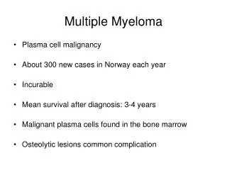
Multiple Myeloma (MM) is “a malignant disorder of the monoclonal plasma cells.” • Is characterized by lytic bone lesions, infiltrate in the bone marrow, hypercalcemia, renal failure, anemia, and a monoclonal protein in the serum or urine2 • Remains an incurable neoplasm of plasma cells that affects more than 20,000 people annually in the United States3 Men and African Americans have twice the incidence of MM as Women and Whites, respectively!!4 Introduction
MM accounts for 1% of all malignancies and is the 2nd most common hematologic malignancy with prevalence of around 10%. • The prevalence of MM was around 64,615 in 2008.1 Epidemiology
Plasma Cells • Pathogenesis • MM represents a stepwise process of malignancy: • Premalignant Stage: Monoclonal gammopathy of undetermined significance (MGUS), with a number of cytogenetic and gene abnormalities1. • Malignant Stage iIncludes: • Induction of angiogenesis3 • Suppression of cell immunity3 • Involvement of cytokines such as IL-6 and VEGF.5 Patho-Biology
Cytogenetic Alterations: • It has been well documented that all MM develops from MGUS, and the initial event required for transformation to MGUS provides the first step.1 • The most common alteration is hyperdiploidy.2 • Additional recurrent abnormalities include loss of chromosome 13.2 Cytogenetics
Growth of the MM Cell in the BM Microenvironment • Myeloma cells interact with BM stromal cells, leading to cytokine production and adhesion signaling changes. • This change affects the migration and localization of the myeloma cells in the BM.1,2 • Moreover, proliferative and antiapoptotic signaling cascades activated in myeloma as a result of these interactions include PI3K/Akt, MAPK kinase (MEKK)/ERK, JAK 2/STAT 3, and NFκB pathways. MM In The Bone Marrow
The progression of MGUS to myeloma is characterized by the development of bone lesions. • The pathogenetic mechanisms involved are complex and involve a combination of osteoclast activation coupled with osteoblast inhibition.2 • There are several important mechanisms that mediate increased osteoclast activation. • There is an increase in NF-κB ligand RANKL expression by osteoblasts and possibly plasma cells.3,4 MM In The Skeleton
The same type of screening is recommended for Male (XY) and Female (XY)1 • Screen for multiple myeloma every 6 to 12 months in patients with a diagnosis of MGUS.1 • Recognize that MGUS precedes symptomatic MM.2 • CBC, Ca++ level, Cr. level, Serum free light chains, and 24-hour urine protein electrophoresis.3 Screening
Ancillary Studies • If the serum M-spike is >1.5 g/dL, obtain a bone survey and a bone-marrow aspirate.1 • If these studies are normal, repeat the serum protein electrophoresis, CBC, creatinine, and calcium at 6 months if <2.0 g/dL and at 3 months if ≥2.0 g/dL.2,3 • If values remain stable, reduce screening frequency to yearly in the <2.0 g/dL group and every 6 months in the ≥2.0 g/dL group.2,3 Screening
A thorough history to look for manifestations of the disease and its complications. • Although the symptoms of MM can be nonspecific they are usually more pronounced in FEMALES.1 • Symptoms of MM can reflect: • Anemia, hypercalcemia, lytic bone lesions, hyperviscosity or thrombocytopenia, soft-tissue plasmacytomas, or hypogammaglobulinemia.2 Diagnosis
*C: Calcium elevation (> 10.5 mg/L or ULN)R: Renal dysfunction (serum creatinine > 2 mg/dL)A: Anemia (Hb < 10 g/dL or 2 g < normal)B: Bone disease (lytic lesions or osteoporosis) Diagnostic Criteria for Myeloma
Use laboratory studies to confirm the diagnosis of MM and differentiate it from other plasma-cell disorders!1,2,3 • Test you must have: • CBC • Serum calcium and creatinine • Serum Protein Electrophoresis • Serum free light chains • Quantitative immunoglobulins • 24-hour urine PEP • Serum β-2 microglobulin • Radiographic bone survey! Lab-Work
Osteolytic Lesion • The most common symptoms are fatigue and bone pain.1 • Osteolytic bone lesions can be detected on routine x-rays, MRI, CT, or combined FDG-PET/CT.2 • Bone pain may present as an area of persistent pain or be migratory. • Often in the lower back and pelvis. • Pain may be sudden in onset when associated with a pathological fracture and is often precipitated by movement.2 A Bone Tests
Performed to quantify the monoclonal proteins present in myeloma.1 • 70% is IgG • 20%, it is IgA • 5% to 10% is light chains only2 • After recognition of a localized band suggestive of an M protein on SPEP, immunofixation is necessary for confirmation and to determine the heavy- and light-chain class of the M protein.3 Protein Electrophoresis
Risk-stratify patients with MM using established staging systems and prognostic factors1 • Use clinical and lab data to stage patients with MM using the Durie-Salmon Staging System or the International Staging System.1,2 • Classify as stage I, II, or III, • Stage I having the most favorable prognosis3 • Understand that important prognostic factors include3,4: • Albumin, Hb, Ca++, β2-microglobulin • M-component production rate • Bone x-ray results| International Staging System Staging – Risk Stratification
First-in-class proteasome inhibitor • Bortezomib • Carfilzomib • Via inhibition of proteasomal degradation of regulatory proteins, it fosters the following • Antiproliferation • Proapoptosis • Antiangiogenesis Proteasome Inhibitors: Mechanism Of Action
Immunomodulators • Thalidomide • Lenalidomide • Pomalidomide • Induction of • Apoptosis • Antiproliferative effects • Inhibition of • Angiogenesis • Tumor necrosis factor • Interleukin-6 • Male and female have similar outcomes Immune Modulating Agents: Mechanism Of Action
Alkylating Agents Melphalan alkylates the DNA nucleotide, guanine, and causes linkages between strands of DNA. This chemical alteration inhibits DNA synthesis and RNA synthesis functions necessary for cells to survive. These changes cause cytotoxicity in both dividing and non-dividing tumor cells.1
Physiological changes during pregnancy affect drug distribution, metabolism, and excretion. • No pharmacokinetic studies have been conducted that aim to assess whether pregnant women should be treated with different chemotherapy doses.1 • Most cytotoxic agents are known to cross the human placenta and reach the fetus circulation • Exposure to chemotherapy during the first trimester has been associated with a 10–20% risk of major congenital malformations and a 33% risk of adverse pregnancy outcomes.2 MM and Pregnancy
Standard remains high dose chemotherapy followed by ASCT for patients with good performance status. • Autologous stem cell transplant has shown to improve survival. • Up-front transplantation with novel drug induction can induce high CR rates and increase quality of life • After short-course induction, with relapse after ASCT, patients can experience response to rescue therapies or retreatment. Autologous Stem Cell Transplantation (ASCT)
Some of the trials fail to provide data on sub group analysis based on Sex and Gender,1,2while the others showed no statistical difference in efficacy of currently practiced therapeutic drugs between males and females.3,4,5 SEX AND GENDER BASED DIFFERENCE IN EFFICACY OF AVAILABLE THERAPEUTIC MODALITIES