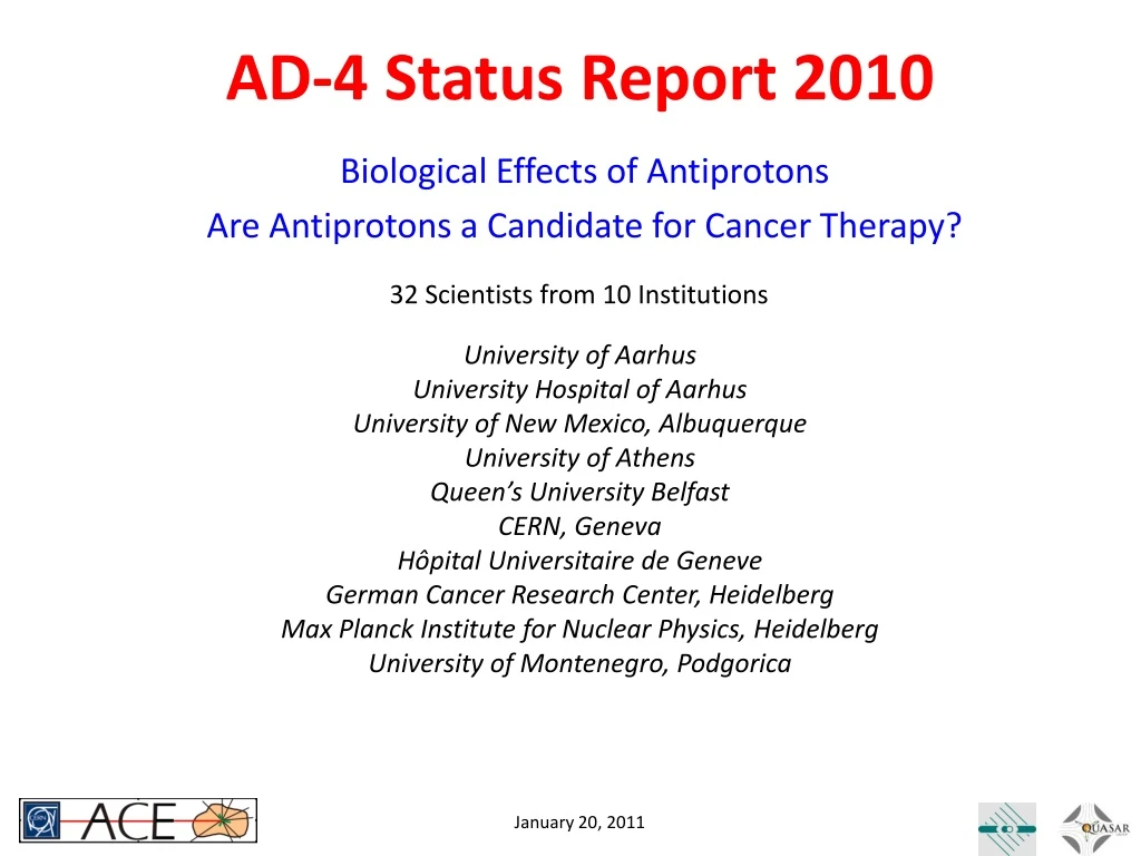
AD-4 Status Report 2010
320 likes | 333 Vues
This status report explores the potential use of antiprotons in cancer therapy, highlighting their physical advantages and potential clinical advantages over other particle types. The report also presents the findings from the AD-4 experiment at CERN, analyzing the biological effects of antiproton irradiation on V-79 Chinese Hamster cells. Additional analysis includes the variation of relative biological effectiveness (RBE) with depth and the use of a new beam monitor for precise dose calculations.

AD-4 Status Report 2010
E N D
Presentation Transcript
AD-4 Status Report 2010 32 Scientists from 10 Institutions University of Aarhus University Hospital of Aarhus University of New Mexico, Albuquerque University of Athens Queen’s University Belfast CERN, Geneva HôpitalUniversitaire de Geneve German Cancer Research Center, Heidelberg Max Planck Institute for Nuclear Physics, Heidelberg University of Montenegro, Podgorica Biological Effects of Antiprotons Are Antiprotons a Candidate for Cancer Therapy?
Rationale for Conformal Radiotherapy Dose (and tumor control) are limited due to tolerance of organs at risk Dose to Target Better conformity of dose to target enables application of higher doses & higher tumor control without increasing normal tissue complication rate
Particle Therapy offersReduced Integral Dose to Body Particles deposit LESS physical dose in front of the tumor and NO dose beyond the distal edge of the Bragg peak!
Potential Clinical Advantages? • Each Particle Type shows distinct features • Protons are well known and easy to plan (RBE = 1) which is the reason they are most widely adopted. • Antiprotons have lowest entrance dose for the price of an extended isotropic low dose halo. • Carbon ions have sharpest lateral penumbra but comparatively higher entrance dose than even protons (no RBE included here), but show forward directed tail due to in beam fragmentation. Detailed dose plans (including RBE) will need to be developed to assess applicability of particle types for different tumor types and locations!
The AD-4 Experiment at CERN • INGREDIENTS: • V-79 Chinese Hamster cells embedded in gelatin • Antiproton beam from AD(126 MeV) • METHOD: • Irradiate cells with dose levels to give survival in the peak is between 0 and 90 % • Slice samples, dissolve gel, incubate cells, and look for number of colonies ANALYSIS: • Study cell survival in peak (tumor) and plateau (skin) and compare the results to protons (and carbon ions)
Biological Analysis Method Co-60 Dose RBEplat = Plateau Dose =1.0 RBEpeak = Co-60 Dose =1.15 Peak Dose • Plot “peak” and “plateau” survival vs. relative dose to extract the Biological Effective Dose Ratio BEDR = F • RBEpeak/RBEplateau (F = ratio of physical dose in peak and plateau region) Plot “peak” or “plateau” survival vs. absolute dose and compare to 60Co irradiation comparing dose values needed for Iso-Effect for peak, plateau, and 60Co irradiation: Relative Biological effectiveness RBE Example: Protons at Triumf
Carbon Ions – SOBP at GSI note: clinical beams with precise dosimetry and fast dose delivery ……..Energy to achieve same clinical relevant depth and form SOBP as at CERN….
RBE for Carbon Ions Extract survival vs. dose plot for each depth slice and calculate RBESF=10% RBEplateau = 1.2 RBEpeak = 2.0 RBE distal = 1.5
RBE Analysis for Antiprotons • Physical Dose Calculations requires exact Knowledge of Beam Parameters • Biological Variability necessitates multiple Independent Experiments under Identical Conditions • Most important result is NOT Peak-to-Plateau ratio but Variation of RBE with Depth
CERN DATA 2008 note: good control over dose planning for SOBP……..RBEplateau= 1.2 RBEpeak= 1.73 – 2.2
CERN DATA 2009 note: attempt to collect data in tail for RBE analysis…….. difficult task – long irradiation times – very little effect
CERN DATA 2010 Additional data set in Plateau and Peak (preliminary analysis) ……detailed dose calculations and error analysis still ongoing
New Beam Monitor • Purpose: Shot-to-shot monitoring of beam spot shape, size, and position for precise dose calculations • to replace Gafchromic film, facilitate alignment, and have instant feed back on beam changes • Solution: Solid state pixel detector (Monolithic Active Pixel Sensor) • Dedicated MAPS design to beam monitoring • pixel 153×153µm2 squares • two 9×9 interdigitedarrays of n-well/p-epi diodes +twoindependentread-out circuits – avoidingdead time • In-pixel storage capacitors – choice ~0.5pF or ~5pF to cope with signal range Mimotera, Massimo Caccia (Universita’ dell’Insumbria Como, Italy) Long term goal: Measure LET distributions in 2D/3D
Complete Info on Beam Shot Integral, Width in X and Yfor each shot at a glance
Beam Shift!! Mimotera allows immediate response to Accelerator Problems Failure of quadrupole was detected and repaired within 1 hour, and 12 hour irradiation of cell samples (half way completed) was saved!
Real Time Imaging - Simulation • Use 4 x 3 layers of (virtual) silicon pixel detectors 40x40 cm2 / 5 cm spacing 30 cm from origin • Pixel Resolution s = 100 mm • Track charged pions and photons • Overall detector efficiencye= 1% • Define vertex as approach of two tracks closer than 2 mm • If more than 2 tracks per particle are detected use meta-center of vertices of any two tracks
Results of Simulation Beam Eye View Side View Achievable Precision: +/- 1mm
Proof of Principle Experiment • Q: How to minimize material and cost • A: Instead of 3 layers use one layer and look at grazing incidence. • Q: What detector to use for first test?A: Turn to our friends in ALICE and use one (spare) module of the Alice Silicon Pixel Detector (SPD)
First Experimental Realization pbar Water phantom • grazing incidence of pions produce long tracks • length distribution changes with angle • stopping distribution along z-axis can be inferred • Future work: Expansion to 3 - D 289.5° Bragg Peak 293.0° distal fall-off π±
First Results red: Simulation blue: Experiment Distal Edge of Depth Dose Profile is detected Resolution is limited due to distance from target and pion scattering
Monte Carlo for Clinical Beams Monte Carlo for Clinical Example: Distance beam to detector = 30 cm Continuous spill 1 x 10⁹ antiprotons (blue: 1x10⁸) Detection of distal edge possible with precision of 1 – 2 millimeter
DNA Damage and Repair • Quantify DNA damage in human cells along and around a 126 MeV antiproton beam at CERN. • Investigateimmediate and longer term DNA damage. • Investigate non-targeted effects outside the beam path due to secondary particles or bystander signaling.
DNA Damage and Repair Assays • There is more to biology than just clonogenics – especially outside the targeted area: • Immediately after attack on DNA proteins are recruited to the site • This event signals cell cycle arrest to allow repair • If damage is too extensive to repair programmed cell death (apoptosis) is induced • Cells also deficient of cell cycle check point proteins may enter mitosis (cancer cells are often deficient in repair proteins and continue dividing) g-H2AX: Phosphorylation of H2AX in the presence of Double Strand Breaks Micronuclei: Fluorescent detection of micronuclei (parts of whole chromosomes) formed due to DNA damage, which are indicating potential of tumorigenesis g-H2AX and Micronucleus assays are typically used to study immediate and long term DNA damage respectively
Results g-H2AX Assay γ-H2AX foci in cells irradiated with up 1.1x10⁹antiprotons in theplateau (blue) or SOBP (red).
Results for g-H2AX SOBP antiprotons generate largerDNA double strand breaks than either plateau antiprotons or X-rays. 60 minutes after radiation no differenceis detected anymore
Results Micronuclei Assay Mean number of micronuclei for two replicate experiments for antiproton plateau and SOBP data sets. Sub lethal damage seems to be LET dependent.
Summary and Outlook Achievements 2010 • Extended data set on Biological Effect of Antiprotons for preliminary dose planning studies • Confinement of RBE Enhancement to Bragg peak only has been confirmed (preliminary analysis) • DNA damage assays for studies of late effects achieved higher resolution • Fast Beam Monitoring implemented • Real Time Imaging of Stopping Distribution – Proof of principle experiment performed
Summary and Outlook Recent Publications • Bassler, N., Holzscheiter, M.H., Petersen, J.B., 'Neutron Fluencein Antiproton Radiotherapy, Measurements and Simulations', submitted to ActaOncologica (2010) • Bassler, N., Kantemiris, I., Karaiskos, P., Engelke, J., Holzscheiter, M.H., Petersen, J.B. 2010; Comparison of optimized single and multifield irradiation plans of antiproton, proton, and ion beams, Radiotherapy & Oncology (2010) vol. 95, pp. 87 – 93 • Kantemiris, I., Angelopoulos, A., Bassler, N., Giokaris, N., Holzscheiter, M., Karaiskos, P., Kalogeropoulos, T.E., 'Real-time imaging duringantiproton radiotherapy',Phys. Med. Biol. (2010) vol. 55, pp. N1–N9 • J.N. Kavanagh, F.J. Currell, D.J. Timson, M.H. Holzscheiter, N. Bassler, R. Herrmann, G. Schettino; ‘Induction of DNA Damage by Antiprotons for a Novel Radiotherapy Approach’;European Physical Journal DD 60 (2010) pp. 209- 214
Summary and Outlook Future Work • Continue increasing precision of RBE determinationAdd more independent data sets (identical conditions)Improving biological protocols and error analysis.Increase data density for sets to stabilize fits • Detailed dose planning studies on specific cancers • Continue DNA damage studies to assess risk of secondary primary malignancies (SPM’s) • Develop 2D LET measuring system (mostly off line) • Transfer technology between HIT and AD-4 - Protons, Carbon ions, Antiprotons
Beam Time Request 2 weeks of 126 MeV (500 MeV/c) antiprotons • Survival measurements on V-79 cell linesAugment or complete data set on RBEImprove error analysis • Study of DNA damage usingg-H2AX and MNIncreasing statistical significance and complete measurement from 2010 (beam time loss) • Liquid ionization chamber measurementsTwo dose rate method on pulsed beams