Health Physics
Health Physics. Revision. Thermometer . Has some physical quantity that changes with temperature, eg. In a laboratory thermometer, the volume of alcohol changes as temperature changes. Resistance of thermistor changes with temperature in an electronic thermometer. Clinical Thermometer.

Health Physics
E N D
Presentation Transcript
Health Physics Revision
Thermometer • Has some physical quantity that changes with temperature, • eg. In a laboratory thermometer, the volume of alcohol changes as temperature changes. • Resistance of thermistor changes with temperature in an electronic thermometer
Clinical Thermometer • Need to know all the steps you take in using a clinical thermometer to measure temperature • Need to know purpose of the kink • Normal body temperature and hypothermia / fever / diagnosis
Sound • Caused by vibrations • Doesn’t travel through vacuum • Travels at different speeds in different materials • Stethoscope uses body sounds to diagnose illness (lungs/ heart)
Stethoscope • Closed bell – lungs – high frequency • Open bell – heart – low frequency • Sounds picked up at bell/chestpiece • Transmitted through the air in the tube • Passed to the ears by way of the earpieces
Hearing • Range: 20Hz – 20kHz • Ultrasound – frequency above the range of human hearing • Noise levels – dB • >90dB – dangerous for long periods • Ear muffs – absorb some of the energy to reduce the noise level at your ears • Normal conversation: 60dB
Ultrasound Scanning • Ultrasound waves sent into body (gel used to cut down reflections at the skin) • Waves reflected by soft tissue and bones • Reflected waves are detected. Can work out how far away objects are using d=vt if you know the speed of the ultrasound waves • Ultrasound good for baby scans – doesn’t harm humans, like x-rays can.
Light • Should know about reflection, refraction and lenses • Should know about eye defects and how to correct them • Lens power
Endosope • Light is shone down an optical fibre into the body. Light source is outside the body so that only light (no heat) travels down – “cold light”. • Light reflects inside patient • Reflected light travels back up a different fibre optic cable (the image guide) • Light travels by total internal reflection.
Electromagnetic Spectrum • Laser – visible light – vaporising tumours, removing tattoos, eye surgery (laser scalpel) • Infra-red – unhealthy tissue hotter than surroundings – thermogram can pick this up • UV – sterilising surgical instruments, treating acne
Electromagnetic Spectrum 2 • X-rays – can cause cancer • Useful for scanning (normal or CT scans) • Also useful for damaging cancer cells
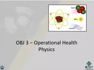
Radioactivity • You should know about alpha, beta, gamma – what they are and how they can be blocked, how strongly ionising they are • GM tube detects radiation using the fact that it causes ionisation – you should be able to explain this • Activity is measured in Becquerels (1 count per second) • You should be able to do half life calculations
Biological Harm • Harm caused by radiation depends on: • Type of tissue • Type of radiation • Total amount of energy • Equivalent dose takes account of biological harm – measured in Sieverts (Sv) • Minimise harm by shielding, distance and measurement
Film Badges • Photographic film is a detector of radiation and x-rays • Can have a badge with film loaded in it with various materials in different windows on the badge – can tell what type of radiation was present by seeing where the film is exposed.
Radiation in Medicine • Scanning: • inject / swallow gamma source • Allow time for source to reach tissue / organ • Detect radiation using gamma camera • Use gamma because it can be detected outside the body • Choose half life long enough for scan to take place but not too long that harm will be done.
Radiation in Medicine • Killing cancer cells • Can use gamma (from outside the body) or alpha (injected / placed inside the body) • Radiation kills cancer cells – also damages surrounding healthy tissue. • This is called radiotherapy