Download
1 / 40
400 likes | 508 Vues
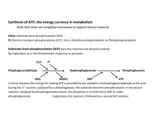
Regulation of the subcellular localization and phosphorylation of the inositol 1,4,5-trisphosphate receptor. 18/06/2004. DAG. α. ß. . IP 3. Ca 2+ signals. Ca 2+. ADP. ATP. PMCA. Ca 2+. General introduction. RTK. PIP 2. PIP 2. Na + /Ca 2+ exchanger. GPCR. PLC ß. PLC . α.

E N D
Regulation of the subcellular localization and phosphorylation of the inositol 1,4,5-trisphosphate receptor 18/06/2004
DAG α ß IP3 Ca2+ signals Ca2+ ADP ATP PMCA Ca2+ General introduction RTK PIP2 PIP2 Na+/Ca2+ exchanger GPCR PLC ß PLC α α ß Gq IP3R Ca2+ ER Ca2+ influx channel SERCA
[Ca2+]i time Ca2+ signals → regulation in time and space: “spatio-temporal character” • elementary signals: • blips • puffs • global signals: • oscillations • waves → cell-type dependent and the underlying mechanisms are still unknown PART ONE:Subcellular localization of IP3Rs PART TWO: Differential regulation of IP3R isoforms
PART ONE: The regulation of the subcellular localization of IP3Rs in A7r5 smooth muscle cells
Overview part one The regulation of the subcellular localization of IP3Rs in A7r5 smooth muscle cells. • Introduction • Subcellular localization of IP3R1 and IP3R3 in A7r5 • Localization of IP3Rs in other cell types • Interaction of IP3Rs with cytoskeletal proteins and protein kinases
Ligand binding regulatory channel 1. Introduction • IP3R = intracellular Ca2+-release channel located predominantly • on ER membranes • 3 known isoforms: IP3R1, IP3R2 and IP3R3
Distribution of IP3Rs dependent on cell type • Different isoforms can have different localizations in the cell • One isoform can have more than one localization in the cell • Short-term agonist stimulation can lead to clustering of IP3Rs → role of long-term stimulation
3 µM AVP 5 h 2. Subcellular localization of IP3R1 and IP3R3 in A7r5 IP3R1 IP3R3 Control
80 cytoplasmic 70 60 50 Cells (%) 40 30 perinuclear 20 10 0 0 1 2 3 4 5 Time (h) Redistribution occurs mostly during the second hour after addition of AVP and the process is reversible Time course of IP3R1 redistribution
100 90 80 70 Relative fluorescence (%) 60 50 40 30 20 80 10 70 0 60 0 1 2 3 4 5 50 Time (h) Time (h) Cells (%) 40 30 20 10 0 Redistribution of IP3R1 = downregulation? Long term stimulation → downregulation of IP3Rs cytoplasmic perinuclear 0 1 2 3 4 5 IP3R1 Kinetics of both processes are completely different.
80 70 60 50 40 30 20 10 0 Downregulation redistribution IP3R1 Downregulation → ubiquitin-proteasome pathway Cells with perinuclear IP3R1 (%) control 3 µM AVP 20 µM MG-132 + 3 µM AVP
Imipramine PLC ß CPA OAG Gö-6976 + AVP Staurosporine + AVP Imipramine thapsigargin Bisindolylmaleimide + AVP Thapsigargin CPA Staurosporine Bisindolylmaleimide Gö-6976 OAG PKC plays an important role in the redistribution of IP3R1 Factors involved in the redistribution of IP3R1 Vasopressin (AVP) DAG PIP2 V1a α PKC α α ß ß Gq IP3 IP3R Ca2+ ER AVP Control SERCA
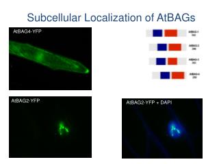
PDI SERCA ER structure seems to be unaffected, but proteins involved in Ca2+ signaling redistribute after prolonged stimulation. Localization of ER proteins ER-targeted EYFP Control AVP
control AVP OAG microtubuli actin Mechanism of redistribution of IP3R1 a) Cytoskeleton
An intact microtubular network is necessary for redistribution of IP3R1
Control AVP 14°C AVP 37°C Brefeldin A Redistribution could occur through vesicle trafficking b) Vesicle trafficking % cells with perinuclear IP3R1
HeLa 16HBE14o- SH-SY5Y IP3R1 IP3R3 IP3R3 IP3R1 untreated untreated untreated Carbachol (10 µM) Histamine (1 µM) ATP (50 µM) TG (1 µM) TG (1 µM) TG (1 µM) Bar = 10 µM OAG (50 µM) 3. Localization of IP3Rs in other cell types
Vimentin Tubulin Zyxin Actin Talin Vinculin Proteins expressed in A7r5: 4. Interaction of IP3Rs with cytoskeletal proteins and protein kinases in A7r5 Restriction of IP3Rs in a specific part of the cell could be due to interactions with cytoskeletal proteins e.g. focal adhesion proteins.
IP3R3 interacts with talin and actin, while IP3R1 only interacts with talin. Immunoprecipitation of IP3R1 and IP3R3 with cytoskeletal proteins
IP3R3 interacts with the regulatory subunit of PKA. Immunoprecipitation of IP3R1 and IP3R3 with protein kinases Heavy chain IP3R3 antibody Negative control beads Pre-immune serum IP IP3R3 IP IP3R1 Lysate PKA PKC Even after stimulation of cells with OAG during different times, no co-immunoprecipitation of IP3R1 or IP3R3 with PKC was found.
SUMMARY PART ONE 1. IP3R1 redistribution in A7r5 cells • IP3R1 and IP3R3 have a different subcellular localization in A7r5 • IP3R1 and SERCA redistribute after prolonged stimulation with AVP • Redistribution downregulation: 2 independent processes • Activation of PKC is sufficient to induce redistribution • ER structure undergoes no major morphological changes • An intact microtubular network is necessary for redistribution • Process likely occurs through vesicular trafficking
2. Interaction of IP3Rs with cytoskeletal proteins and protein kinases • IP3R3 interacts with actin and talin, while IP3R1 only interacts • with talin • IP3R3 interacts with the regulatory subunit of PKA
PART TWO: The regulation of the phosphorylation of IP3R1 and IP3R3 by PKC
Overview part two The regulation of the phosphorylation of IP3R1 and IP3R3 by PKC. • Introduction • In vitro phosphorylation of IP3R1 and IP3R3 by PKC • Determination of the phosphorylation site(s) on IP3R1 and IP3R3 • Influence of PKA phosphorylation on PKC phosphorylation • Differential regulation of the PKC phosphorylation by Ca2+ and CaM • Potential role in the negative slope of the bell-shaped effect of Ca2+
IP3 IP3R1, IP3R2, IP3R3 IP3R1, IP3R2 CaM Stimulation of IICR ≤ 300 nM ≥ 300 nM [Ca2+]i 1.Introduction Regulation of IP3R activity Ligand binding domain Regulatory domain Channel domain * Ca2+ * CaM → Nael
1.Introduction 1.Introduction Regulation of IP3R activity Regulation of IP3R activity Ligand binding domain Ligand binding domain Regulatory domain Regulatory domain Channel domain Channel domain * Ca2+ * CaM → Nael IP3 IP3 * ATP … * contain several consensus sequences for different kinases.
IP3R1: - PKA - PKG - PKC - CaMKII - Fyn and Lyn - Rho kinase - cdc2/cyclin B - ERK IP3R2: - PKA - Rho kinase IP3R3: - PKA - Rho kinase stimulates IICR ? stimulates IICR? ? stimulates IICR stimulates IICR IP3 binding ↑ ? ? no effect on IICR? ? no effect on IICR?
2. In vitro phosphorylation of IP3R1 and IP3R3 by PKC Catalytic subunit of PKC (, , ) IP3Rs purified from Sf9 microsomes on sucrose density gradient and/or CaM-sepharose column → highly purified fraction. IP3R1 32P phospho (Ser) PKC Ab
Autoradiography Rbt475 IP3R3 is also phosphorylated, but to a lesser degree
3. Determination of the phosphorylation site(s) on IP3R1 and IP3R3 →controlled proteolysis with -chymotrypsin after in vitro phosph. IP3R1 Yoshikawa et. al., J. Biol. Chem., 1999, 274, 316-327 5 fragments
0.2 µg/ml CT 0.4 µg/ml CT Control 273 kDa 225 kDa 210 kDa 130 kDa 40 kDa Phosphorylated fragments
3. Determination of the phosphorylation site(s) on IP3R1 and IP3R3 →controlled proteolysis with -chymotrypsin after in vitro phosph. IP3R1 Yoshikawa et. al., J. Biol. Chem., 1999, 274, 316-327 Anti-(1829-1848) 5 fragments
0.2 µg/ml CT 0.4 µg/ml CT Control 273 kDa 273 kDa 225 kDa 210 kDa 225 kDa 210 kDa 130 kDa 130 kDa Blot stripped and reprobed 40 kDa 40 kDa Phosphorylated fragments anti-(1829-1848) Ab
IVa 40k
S1852? IP3R1 S1832? IP3R3 CaCaM mass spectrometry: IP3R1 and IP3R3 performed by Prof. E. Waelkens (Biochemie) • Phosphorylated receptor → SDS-PAGE • In gel digestion • Extraction of proteolytic peptides • Concentration of phosphopeptides • Mass spectrometry
Prior phosphorylation with PKA enhances PKC phosphorylation Pathways activating PKA and PKC may have synergistic effects on the potentiation of Ca2+ release by IP3R1. 4. Influence of PKA phosphorylation on PKC phosphorylation IP3R1
+ Ca2+ (50 µM) - Ca2+ IP3R1 IP3R3 IC50 ≤ 2 µM Ca2+ inhibits PKC phosphorylation 5. Differential regulation of the PKC phosphorylation by Ca2+ and CaM Ca2+
CaM CaM1234 CaM1234 CaM inhibits the phosphorylation of IP3R1 CaM
CaM Ca2+-dependent kinases and phosphatases may play an important role in the regulation of the IP3R. 6. Potential role in the negative slope of the bell-shaped effect of Ca2+ Stimulation of IICR ≤ 300 nM ≥ 300 nM [Ca2+]i
SUMMARY PART TWO • Both IP3R1 and IP3R3 are in vitro phosphorylated by PKC, although • to a different extent. • IP3R1 contains 1 PKC phosphorylation site, probably S1852. • IP3R3 also contains 1 PKC phosphorylation site, probably S1832. • Prior phosphorylation of IP3R1 with PKA enhances PKC-mediated • phosphorylation → pathways involving PKA and PKC may have a • synergistic effect. • Ca2+ and CaM inhibit PKC phosphorylation of IP3R1 • → could play a role in the negative feedback of • Ca2+ and CaM on IP3R function. • IP3R3 phosphorylation is not affected by Ca2+ and CaM.