Fundoscopy
Fundoscopy. Using an ophthalmoscope. Objectives. Identify patients at risk from eye disease Recognising the normal fundus Describe Patient preparation Demonstrate correct procedure using the ophthalmoscope Describe abnormal findings, and recognise disease process .

Fundoscopy
E N D
Presentation Transcript
Fundoscopy Using an ophthalmoscope
Objectives • Identify patients at risk from eye disease • Recognising the normal fundus • Describe Patient preparation • Demonstrate correct procedure using the ophthalmoscope • Describe abnormal findings, and recognise disease process
Chronic diseases(HIV arteriosclerosis) Diabetes Hypertension CVA Cardiac disease Pts on certain drugs (anticholinergics / steroids Top 5 Common eye conditions Cataract Glaucoma Age related macular degeneration Squint Diabetic eye disease Patients requiring ophthalmologyinvestigation
Normal fundus • Disc: Clear outline; optic cup is pale and centrally located. • Retina: Normal red/orange colour, macula is dark. The macula is approximately 2 disc diameters away from disc and 1.5 degrees below horizon. • Vessels: Arterial/venous ratio 2 to 3; the arteries appear a bright red, the veins a slightly purplish colour.
What to observe • Optic disc- colour/size/edges • Cup – size • Blood vessels – number/width/tortuosity • Macular / fovea • Other findings –haemorrhages, soft and hard exudates, oedema
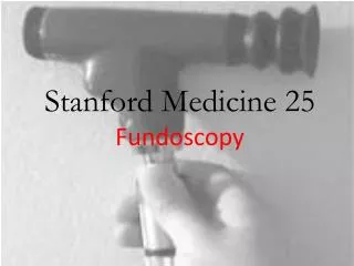
Procedure • Ask patient to fix stare at object • Turn on scope and set dial to 0 • Remember right eye right hand • Rest hand on patients forehead • Begin at arms length away, should see red reflex. • Move closer until optic disc visible ( aprox. 3-5cms) Turn dial until disc in focus. • Hyperopic – turn disc for plus numbers (green) • Myopic – minus numbers (red)