Molecular Graphics Perspective of Protein Structure and Function
250 likes | 483 Vues
Molecular Graphics Perspective of Protein Structure and Function. VMD Highlights. > 20,000 registered Users Platforms: Unix (16 builds) Windows MacOS X Display of large biomolecules and simulation trajectories Sequence browsing and structure highlighting

Molecular Graphics Perspective of Protein Structure and Function
E N D
Presentation Transcript
Molecular Graphics Perspective of Protein Structure and Function
VMD Highlights • > 20,000 registered Users • Platforms: • Unix (16 builds) • Windows • MacOS X • Display of large biomolecules and simulation trajectories • Sequence browsing and structure highlighting • User-extensible scripting interfaces for analysis and customization
VMD Permits Large Scale Visualization Purple Membrane 150,000 Atoms • Large structures: 300,000 atoms and up • Complex representations • Long trajectories: thousands of timesteps • Volumetric data • Multi-gigabyte data sets break 32-bit barriers • GlpF: each 5 ns simulation of 100K atoms produces a 12GB trajectory F1 ATPase 327,000 Atoms
Focus on two proteins Ubiquitin Bovine Pancreatic Trypsin Inhibitor (BPTI) Ubiquitin BPTI
Ubiquitin • 76 amino acids • highly conserved • Covalently attaches to proteins and tags them for degradation
Glycine at C-terminal attaches to the Lysine on the protein by an isopeptide bond. • it can attach to other ubiquitin molecules and make a polyubiquitin chain. • There are 7 conserved lysine residues • on the ubiquitin. 2 ubiquitins attached together through LYS 48. LYS 63 and LYS 29 are also shown there.
Ubiquitination Pathway • Activation by E1 (ATP dependent process) (thiol-ester linkage between a specific cysteine residue of E1 and Glycine on ubiquitin) • Transfers to a cysteine residue on E2 (ubiquitin conjugation enzyme) • E3 transfers the ubiquitin to the substrate lysine residue. • E3 recognizes the ubiquitination signal of the protein.
Ubiquitin Functions: • Tagging proteins to be degraded in proteasome. • degrading misfolded proteins • regulates key cellular processes such as cell division, gene expression, ... • A chain of at least 4 ubiquitins is needed to be recognized by the proteasome.
Independent of proteasome degradation • Traffic Controller • Directing the traffic in the cell. determines where the newly synthesized proteins should go • Tagging membrane proteins for internalization Hicke, L., Protein regulation by monoubiquitin, Nat. rev. mol cell biol., 2, 195-201 (2001)
Regulating gene expression: • (indirectly, by destruction of some of the involved proteins) • Recruiting Transcription Factors(proteins needed for gene expression) • Conformational changes in Histone, necessary before gene expression Hicke, L., Protein regulation by monoubiquitin, Nat. rev. mol cell biol., 2, 195-201 (2001)
Different types of ubiquitin signals • Length of the ubiquitin chain. • How they are attached together. • Where it happens. • multi-ubiquitin chains, linked through Lysine 48, target protein for proteasome degradation. • K63 linkages are important for DNA repair and other functions.
Monoubiquitylation versus multi-ubiquitylation Marx, J., Ubiquitin lives up its name, Science 297, 1792-1794 (2002)
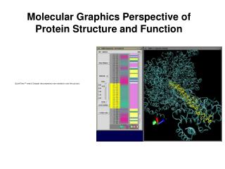
Basics of VMD Loading a Molecule New Molecule Molecule file browser Browse Load
Basics of VMD Rendering a Molecule Current graphical representation Selected Atoms Draw style Coloring Drawing method Resolution, Thickness
Basics of VMD Change rendering style CPK tube cartoon
Basics of VMD Create Representation Delete Representation Current Representation Material Multiple representations
VMD Scripting Left: Initial and final states of ubiquitin after spatial alignment Right (top): Color coding of deviation between initial and final
VMD Sequence Window Beta Value List of the residues Structure T: Turn E: Extended conformation H: Helix B: Isolated Bridge G: 3-10 helix I: Phi helix Zoom
VMD Macros to Color Beta Strands Use VMD scripting features to color beta strands separately; show hydrogen bonds to monitor the mechanical stability of ubiquitin Ubiquitin stretched between the C terminus and K48 does not fully extend!
Discovering the Mechanical Properties of Ubiquitin Ubiquitin stretched between the C and the N termini extends fully!
Discover BPTI on your own! bovine pancreatic trypsin inhibitor • Small (58 amino acids) • rigid • Binds as an inhibitor to Trypsin • (a serine proteolytic enzyme, that appears in digestive system of mammalians.) • Blocks its active site.
Mechanism of cleavage of peptides with serine proteases. Radisky E. and Koshland D. Jr., Proc. Natl. Acad. Sci., USA, 99, 10316-10321 Trypsin: A proteolytic enzyme that hydrolyzes peptide bonds on the carboxyl side of Arg or Lys.
BPTI: A “standard mechanism” inhibitor • Binds to Trypsin as a substrate. (has a reactive site) • forms an acyl-enzyme intermediate rapidly. • Very little structural changes in Trypsin or BPTI. • several H-bonds between backbone of the two proteins • little reduction in conformational entropy binds tightly • Remains uncleaved. • (hydrolysis is 1011 times slower than other substrates) • Structures of the protease binding region, in the proteins of all 18 families of standard mechanism inhibitors are similar.
Why does Trypsin cleave BPTI so slowly? • Disruption of the non-covalent bonds in the tightly bonded enzyme-inhibitor complex, increases the energy of transition states for bond cleavage. • Water molecules do not have access to the active site, because of the tight binding of Trypsin and BPTI. • After the cleavage of the active-site peptide bond, the newly formed termini are held in close proximity, favoring reformation of the peptide bond. • The rigidity of BPTI may also contribute by not allowing necessary atomic motions.