Download
1 / 44
440 likes | 471 Vues
Examining the evolutionary history and function of gene duplication leading to the origins of novel functions. Learn about the interaction of mutations, gene subfunctionalization, and sources of genetic variation, using steroid hormone receptors as examples. Explore how steroid receptors evolved, their roles in signal transduction, and the specificity of ligand binding across different species. Discover the evolutionary journey of aldosterone-mineralocorticoid receptor complex and its implications for understanding molecular system evolution.

E N D
Lesson Today Evolutionary History of Function Gene Duplication leading to evolutionary origins of novel functions How mutations interact to modify function
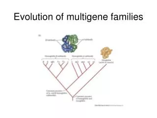
Evolution of novelty Gene Duplication and Subfunctionalization: Examples: Receptors, Enzymes, Developmental genes, etc.: • Hox clusters • Osmoregulatory ion uptake enzymes (ATPases) • Cytp450s (detoxification enzymes) • Olfactory genes • Opsin genes • Hemoglobin
Gene Duplications • Main source of novel genes
Sources of Genetic Variation (type of mutation) • Gene duplications, followed by differentiation • End up with “gene family”: different opsin genes, hemoglobin, ATPases, etc.
Evolution of new functions (and genes) via gene duplications New function Partitioning of function Loss of function
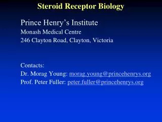
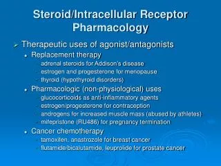
What are Steroid Receptors? I am using this as an example to facilitate understanding of the impacts of pesticides and other environmental toxins on animal physiology (next lectures)
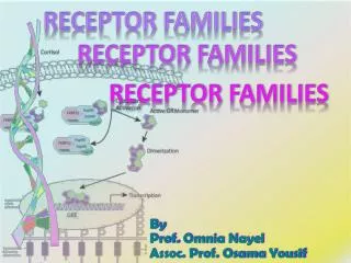
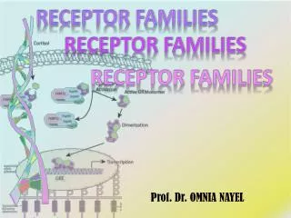
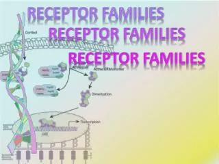
Steroid Hormone Receptors • Transcription factors • Intracellular receptors (typically cytoplasmic) that bind to ligands (e.g. steroid hormones) • Initiate signal transduction which lead to changes in gene expression
Steroid hormones are lipid soluble, bind to cytoplasmic Steroid Hormone Receptors and then enter the nucleus, leading to transcription
The estrogen receptor is • fairly nonspecific, • It is ancestral (phylogeny on next slide), and ancestral receptors tend to be less specific (specificity evolves) • It needs to bind to multiple ligands, ~12 estrogens (estradiol, estriol, estrone, etc.) • So... many compounds will bind to it, such as pesticides Estrogen receptor alpha ligand-binding domain complexed to estradiol
Glucocorticoid receptor Baker, ME. 2001. Adrenal and sex steroid receptor evolution: environmental implications. Journal of Molecular Endocrinology 26:119–125 Estrogen Receptors Mineralocorticoid receptor Sex steroid response probably occurred in the early Cambrian Progesterone receptor Estrogen response evolved in jawless fish or tunicates (early chordates) Androgen receptor Eel ERb Human ERb Trout ERa Xenopus ERa the most ancient of the adrenal and sex steroid receptors Human ERa
How would an integrated molecular system evolve, such as the functional interaction between a hormone and receptor? For example, how could a hormone evolve if a receptor is not present, and visa versa?
Example • Evolution of function of the aldosterone - Mineralocorticoid Receptor (MR) complex • How did this ligand-receptor relationship evolve?
Aldosterone is thought to be a recently derived hormone, and a tetrapod specific hormone (vertebrates with four feet), absent in more anciently derived species
Mineralocorticoid receptor (MR) and the Glucocorticoid receptor (GR) descend from a gene duplication deep in the vertebrate lineage (~450+ mya) and now have distinct signaling functions In most vertebrates, GR is activated by the stress hormone cortisol to regulate metabolism, inflammation, and immunity MR is activated by aldosterone to regulate reabsorption of ions and water and secretion of potassium in the kidneys. MR can also be activated by cortisol
The gene duplication event leading to MR and GR occurred >450 million yrs ago
Background • Functional assays indicate that the ancestral (basal) receptors are activated by very low doses of aldosterone, cortisol, and 11-deoxycorticosterone (DOC); they are similar in this respect to MRs of tetrapods and teleosts (Fig. 2 -next slide) • The only receptors insensitive to aldosterone are the GRs of tetrapods and teleosts • Given these results, the most parsimonious scenario is that AncCR was capable of being activated by aldosterone and that aldosterone sensitivity was lost in the GRs of bony vertebrates (see Fig. 1)
How might have the aldosterone-MR partnership have evolved? If the hormone is not yet present, how could selection drive the receptor’s affinity for it? Conversely, without the receptor, what selection pressure could guide the evolution of the ligand?
Test Hypothesis: • Performed gene resurrection to experimentally examine the function of the ancestral corticoid receptor (AncCR) • Inferred the maximum likelihood (ML) amino acid sequence of AncCR’s ligand-binding domain (see Fig. 1) • Synthesized the AncCR-LBD sequence and expressed it in cultured cells; using a reporter assay
Results AncCR is a sensitive and effective aldosterone receptor (Fig. 3A) Like the extant CRs and MRs, it is also activated by low doses of DOC and, to a lesser extent, cortisol (Fig. 3A) • This result is surprising, because aldosterone has long been considered a tetrapod-specific hormone • Aldosterone is absent from the plasma of lamprey and hagfish (more ancient vertebrates) (Fig. 3B)
WHY would the ancient corticoid receptor respond to a not yet existing hormone (aldosterone)? And how would the specificity between MR and aldosterone evolve?
Fig. 4. Evolution of specific aldosterone-MR signaling by molecular exploitation. (A) Synthesis pathway for corticosteroid hormones. Ligands for the ancestral CR and extant MRs are underlined; cortisol, the ligand for the tetrapod GR, is overlined. The terminal addition of aldosterone is in green. Asterisks, steps catalyzed by the cytochrome P-450 11b-hydroxylase enzyme; only the tetrapod enzyme can catalyze the step marked with a green asterisk. (B) MR’s aldosterone sensitivity preceded the emergence of the hormone. The vertebrate ancestor did not synthesize aldosterone (dotted circle), but it did produce other corticosteroids (filled circle); it had a single receptor with affinity for both classes of ligand. A gene duplication (blue) produced separate GR and MR. Two changes in GR’s sequence (red) abolished aldosterone activation but maintained cortisol sensitivity [see (C)]. In tetrapods, synthesis of aldosterone emerged due to modification of cytochrome P-450 11b-hydroxylase. mya, million years ago. (C) Mechanistic basis for loss of aldosterone sensitivity in the GRs. Phylogenetically diagnostic amino acid changes that occurred during GR evolution were introduced into AncCR-LBD by mutagenesis. Dose-response is shown for aldosterone (green), DOC (blue), and cortisol (red). The double mutant (bottom right) has a GR-like phenotype. Arrows shows evolutionary paths via a nonfunctional (red) or functional (green) intermediate.
Extant MRs retain the ancestral phenotype, so the specificity of the MR-aldosterone relationship is actually due to the secondary loss of aldosterone sensitivity in the GR (Fig. 4B), rather than evolution of specificity for MR.
Which Mutations? • Explored which sequence changes are on the branch where aldosterone sensitivity was lost • Introduced all four single GR-diagnostic states and all six two-fold combinations into AncCR-LBD using mutagenesis and determined their effect on receptor function
Which Mutations? • Replacement of Serine106 with Proline (S106P) and Leucine111 with Glutamine (L111Q) conferred a GR-like phenotype
L111Q alone radically reduces activation by all ligands tested• S106P reduces aldosterone (green) and cortisol (red) sensitivity, but this receptor remains highly DOC-sensitive (blue)• In the S106P background, L111Q further reduces aldosterone sensitivity but now restores cortisol response to levels characteristic of extant GRs When each mutation was introduced in isolation, it was discovered that both are required to yield the GR phenotype
But now, let’s look more closely at the actual transition where the mutations occur • These substitutions recapitulate a large portion of the functional shift from AncGR1 to AncGR2 (~420 to 440 Ma), radically reducing aldosterone and DOC response while maintaining moderate sensitivity to cortisol (Fig. 2A)
Instead of using the ancestral AncCR, the structures of AncGR1and AncGR2 were compared to determine the mechanism by which these two substitutions shift function Ancient GR1 and GR2 were reconstructed using homology modeling and energy minimization based on the AncCR and human GR crystal structures
Fig. 2. Mechanism for switching AncGR1’s ligand preference from aldosterone to cortisol. (A) Effect of substitutions S106P and L111Q on the resurrected AncGR1’s response to hormones. Dashed lines indicate sensitivity to aldosterone (green), cortisol (purple), and DOC (orange) as the EC50 for reporter gene activation. Green arrow shows probable pathway through a functional intermediate; red arrow, intermediate with radically reduced sensitivity to all hormones. (B) Structural change conferring new ligand specificity. Backbones of helices 6 and 7 from AncGR1 (green) and AncGR2 (yellow) in complex with cortisol are superimposed. Substitution S106P induces a kink in the interhelical loop of AncGR2, repositioning sites 106 and 111 (arrows). In this background, L111Q forms a new hydrogen bond with cortisol’s unique C17-hydroxyl (dotted red line).
The major structural difference between AncGR1 and AncGR2 involves Helix 7 and the loop preceding it, which contain S106P and L111Q and form part of the ligand pocket (Fig. 2B). In AncGR1 and AncCR, the loop’s position is stabilized by a hydrogen bond between Ser106 and the backbone carbonyl of Met103.
The movement of helix 7 dramatically repositions site 111, bringing it close to the ligand In this conformational background, L111Q (leucine to glutamine) generates a hydrogen bond with cortisol’s C17-hydroxyl, stabilizing the receptor-hormone complex. Aldosterone and DOC lack this hydroxyl, so the new bond is cortisol specific Replacing Ser106 with proline in the derived GRs breaks this H bond and introduces a sharp kink into the backbone, which pulls the loop downward, repositioning and partially unwinding helix 7
The two substitutions destabilize the receptor complex with aldosterone or DOC Achieves stability with cortisol, switching preference to that hormone This mode of structural evolution is termed “conformational epistasis” because one substitution remodels the protein backbone and repositions a second site, changing the functional effect of substitution at the second site
Fig. 3. Permissive substitutions in the evolution of receptor specificity. (A) Effects of various combinations of historical substitutions on AncGR1’s transcriptional activity and hormonesensitivity in a reporter gene assay. Group Y (L29M, F98I, and S212D) abolishes receptor activity unless groups X (S106P, L111Q) and Z (N26T and Q105L) are present; the XYZ combination yields complete cortisol-specificity. The 95% confidence interval for each EC50 is in parentheses. Dash, no activation. (B) Structural prediction of permissive substitutions. Models of AncGR1 (green) and AncGR2 (yellow) are shown with cortisol. Group X and Y substitutions (circles and rectangles) yield new interactions with the C17-hydroxyl of cortisol (purple) but destabilize receptor regions required for activation. Group Z (underlined) imparts additional stability to the destabilized regions. (C) Restricted evolutionary paths through sequence space. The corners of the cube represent states for residue sets X, Y, and Z. Edges represent pathways from the ancestral sequence (AncGR1) to the cortisol-specific combination (+XYZ). Filled circles at vertices show sensitivity to aldosterone (green), DOC (orange), and cortisol (purple); empty circles, no activation. Red octagons, paths through nonfunctional intermediates; arrows, paths through functional intermediates with no change (white) or switched ligand preference (green).
Evolutionary trajectories that pass through functional intermediates are more likely than those involving nonfunctional steps, so the only historically likely pathways to AncGR2 are those in which the permissive substitutions of group Z and the large-effect mutations of group X occurred before group Y was complete (Fig. 3C). Permissive substitutions stabilized specific structural elements, allowing them to tolerate later destabilizing mutations that conferred a new function
Fig. 4. Structural identification of an ancient permissive substitution. (A) Comparison of the structures of AncCR (blue) and AncGR2 (yellow). Y27R generates a novel cation-p interaction in AncGR2 (dotted cyan line), replacing the weaker ancestral hydrogen bond (dotted red) and imparting additional stability to helix 3. (B) Y27R is permissive for the substitutions that confer GR function. Reporter gene activation by AncGR1 + XYZ (upper right) is abolished when Y27R is reversed (lower right). (Left) Y27R has negligible effect in the AncCR background (or in AncGR1, fig. S9). Green, orange, and purple lines show aldosterone, DOC, and cortisol responses, respectively. Green arrows, likely pathway through functional intermediates.
Evolution of specificity of function • Structural studies of human GR have shown that these two residues change the architecture of the ligand-binding pocket and alter contacts with steroid in ways that exclude aldosterone and facilitate cortisol activation • Results indicate that aldosterone specificity of MR arose from two crucial Amino Acid replacements in the GRs that wiped out ancestral sensitivity to aldosterone • These changes result in evolution of a more specific endocrine response, allowing electrolyte homeostasis to be controlled without also triggering the GR stress response
Molecular Exploitation Functional interaction between aldosterone and mineralocorticoid receptor evolved by a stepwise selective process Ancestral gene resurrection demonstrates that long before the hormone evolved, the receptor’s affinity for aldosterone was present due to its similarity to more ancient ligands (probably DOC) Two amino acid changes in the ancestral sequence resulted in the evolution of present-day receptor specificity Results indicate that tight interactions could evolve by molecular exploitation—recruitment of an older molecule, previously constrained for a different role, into a new functional complex