Download
1 / 109
1.09k likes | 1.23k Vues
Amino acids play a vital role in energy generation through oxidative degradation. This article explores the metabolic conditions that lead to amino acid oxidation, urea production, and the digestion and absorption of dietary proteins. The role of key enzymes and cofactors involved in the process is highlighted.

E N D
Amino Acid Oxidation and The Production of Urea Dr. Fayez Almabhouh Assistant Professor, Biology and Biotechnology Department
Introduction • Amino acids, the final class of biomolecules that, through their oxidative degradation, make a significant contribution to the generation of metabolic energy. • The fraction of metabolic energy obtained from amino acids, whether they are derived from dietary protein or from tissue protein, varies greatly with the type of organism and with metabolic conditions.
Carnivores can obtain up to 90% of their energy requirements from amino acid oxidation, whereas herbivores may fill only a small fraction of their energy needs by this route. • Most microorganisms can scavenge amino acids from their environment and use them as fuel when required by metabolic conditions. • Plants, however, rarely if ever oxidize amino acids to provide energy
In animals, amino acids undergo oxidative degradation in three different metabolic circumstances: • During the normal synthesis and degradation of cellular proteins, some amino acids that are released from protein breakdown and are not needed for new protein synthesis undergo oxidative degradation. • When a diet is rich in protein and the ingested amino acids exceed the body’s needs for protein synthesis, the surplus is catabolized; amino acids cannot be stored. • During starvation or in uncontrolled diabetes mellitus, when carbohydrates are either unavailable or not properly utilized, cellular proteins are used as fuel.
Under all these metabolic conditions, amino acids lose their amino groups to form α-keto acids, the “carbon skeletons” of amino acids. • The α-keto acids undergo oxidation to CO2 and H2O or, often more importantly, provide three- and four-carbon units that can be converted by gluconeogenesis into glucose, the fuel for brain, skeletal muscle, and other tissues.
The Digestion and Absorption of Dietary Proteins Entry of dietary protein into the stomach stimulates the gastric mucosa to secrete the hormone gastrin stimulates the secretion of HCl by the parietal cells and pepsinogen by the chief cells of the gastric glands HCl act as an antiseptic, killing most bacteria and other foreign cells, and a denaturing agent, in addition Pepsinogen converted to pepsin (active) Pepsin hydrolyzes ingested proteins at peptide bonds on the amino-terminal side of the aromatic amino acid residues Phe, Trp, and Tyr acidic stomach contents pass into the small intestine, the low pH triggers secretion of the hormone secretin into the blood.
Secretin stimulates the pancreas to secrete HCO-3into the small intestine to neutralize the gastric HCl, increasing the pH to 7. Releasing of Hormone cholecystokinin, stimulates secretion of several pancreatic enzymes with activity optima at pH 7 to 8. Trypsinogen, chymotrypsinogen, and procarboxypeptidases A and B, the zymogens of trypsin, chymotrypsin, and carboxypeptidases A and B, are synthesized and secreted by the exocrine cells of the pancreas free amino acids is transported into the epithelial cells lining the small intestine enter the blood and travel to the liver
Pyridoxal Phosphate Participates in the Transfer of α-Amino Groups to α-Ketoglutarate • The first step in the catabolism of most L-amino acids, once they have reached the liver, is removal of the α-amino groups, promoted by enzymes called aminotransferases or transaminases.
The effect of transamination reactions is to collect the amino groups from many different amino acids in the form of L-glutamate. • The glutamate then functions as an amino group donor for biosynthetic pathways or for excretion pathways that lead to the elimination of nitrogenous waste products.
Cells contain different types of aminotransferases. • Many are specific for α-ketoglutarate as the amino group acceptor but differ in their specificity for the L-amino acid. • All have the same prosthetic group and the same reaction mechanism. The prosthetic group is pyridoxal phosphate (PLP), the coenzyme form of pyridoxine, or vitamin B6. • PLP functions as an intermediate carrier of amino groups at the active site of aminotransferases.
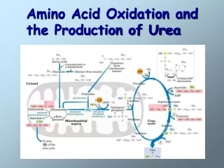
Glutamate Releases Its Amino Group as Ammonia in the Liver • The α-amino acids are collected in the liver in the form of the amino group of L-glutamate molecules • These amino groups must next be removed from glutamate to prepare them for excretion. • In hepatocytes, glutamate is transported from the cytosol into mitochondria, where it undergoes oxidative deamination catalyzed by L-glutamate dehydrogenase.
Reaction catalyzed by glutamate dehydrogenase. The glutamate dehydrogenase of mammalian liver has the unusual capacity to use either NAD+ or NADP+ as cofactor. The mammalian enzyme is allosterically regulated by ADP and GTP . Ammonia (NH+4) produced by these deamination processes
Glutamine Transports Ammonia in the Bloodstream • Ammoniais quite toxic to animal tissues, and the levels present in blood are regulated. • In many tissues, including the brain, some processes such as nucleotide degradation generate free ammonia. • In most animals much of the free ammoniais converted to a nontoxic compound before export from the extrahepatictissues into the blood and transport to the liver or kidneys.
The free ammonia produced in tissues is combined with glutamate to yield glutamine by the action of glutamine synthetase. • This reaction requires ATP and occurs in two steps:
First, glutamate and ATP react to form ADP and a γ –glutamyl phosphate intermediate, which then reacts with ammonia to produce glutamine and inorganic phosphate. • a process catalyzed by glutamine synthetase. • Glutamineis a nontoxic transport form of ammonia; it is normally present in blood in much higher concentrations than other amino acids. • Glutaminealso serves as a source of amino groups in a variety of biosynthetic reactions.
In most terrestrial animals, glutaminein excess of that required for biosynthesis is transported in the bloodto the intestine, liver, and kidneys for processing.
In liver, the amide nitrogen is released as ammonium ion in the mitochondria, where the enzyme glutaminaseconverts glutamine to glutamate and NH+4. In the liver, the ammonia from all sources is disposed of by urea synthesis
Alanine Transports Ammonia from Skeletal Muscles to the Liver • Alanine also plays a special role in transporting amino groups to the liver in a nontoxic form, via a pathway called the Glucose-alanine cycle.
In muscle, glutamate can transfer its α-amino group to pyruvate, a readily available product of muscle glycolysis, by the action of alanine aminotransferase. • The alanine so formed passes into the blood and travels to the liver. Glucose-alanine cycle. • In the cytosol of hepatocytes, alanine aminotransferase transfers the amino group from alanine to α-ketoglutarate, forming pyruvate and glutamate.
Glutamate can then enter mitochondria, where the glutamate dehydrogenase reaction releases NH+4, or can undergo transamination with oxaloacetate to form aspartate, another nitrogen donor in urea synthesis.
Assays for Tissue Damage • Analyses of certain enzyme activities in blood serum give valuable diagnostic information for a number of disease conditions. • Alanine aminotransferase (ALT; also called glutamate-pyruvate transaminase, GPT) and aspartate aminotransferase (AST; also called glutamate-oxaloacetate transaminase, GOT) are important in the diagnosis of heart and liver damage caused by heart attack, drug toxicity, or infection.
After a heart attack, a variety of enzymes, including these aminotransferases, leak from the injured heart cells into the bloodstream. • Measurements of the blood serum concentrations of the GPT, GOT& another enzyme,Creatine kinase (CK) can provide information about the severity of the damage.
The SGOT and SGPT tests are also important in occupational medicine, to determine whether people exposed to carbon tetrachloride, chloroform, or other industrial solvents have suffered liver damage. • Liver degeneration caused by these solvents is accompanied by leakage of various enzymes from injured hepatocytes into the blood.
Aminotransferases are most useful in the monitoring of people exposed to these chemicals, because these enzyme activities are high in liver and can be detected in very small amounts.
Nitrogen Excretion and the Urea Cycle • If not reused for the synthesis of new amino acids or other nitrogenous products, amino groups are channeled into a single excretory end product.
Most aquatic species, such as the bony fishes, are ammonotelic, excreting amino nitrogen as ammonia. • Most terrestrial animals are ureotelic, excreting amino nitrogen in the form of urea. • Birdsand reptilesare uricotelic, excreting amino nitrogen as uric acid. • Plants recycle virtually all amino groups, and nitrogen excretion occurs only under very unusual circumstances
In Ureotelic organisms, the ammonia deposited in the mitochondria of hepatocytesis converted to urea in the urea cycle. • This pathway was discovered in 1932 by Hans Krebs (who later also discovered the citric acid cycle) and a medical student associate, Kurt Henseleit.
Urea production occurs almost exclusively in the liverand is the fate of most of the ammonia channeled there. • The urea passes into the bloodstream and thus to the kidneys and is excreted into the urine
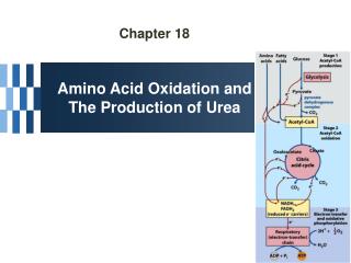
The urea cycle begins inside liver mitochondria, but three of the subsequent steps take place in the cytosol; the cycle thus spans two cellular compartments.
The NH4+generated in liver mitochondria is immediately used,coupling with CO2 as HCO3-to form carbamoyl phosphate. The synthesis of carbamoyl phosphate, through a simple molecule, is complex, comprising three steps, all catalyzed by carbamoyl phosphate synthetase I, this ATP-dependent reaction.
The carbamoyl phosphate, which functions as an activated carbamoyl group donor, now enters the urea cycle. The cycle has four enzymatic steps.
First, carbamoyl phosphate donates its carbamoyl group to ornithine to form citrulline, with the release of Pi. Ornithine plays a role resembling that of oxaloacetate in the citric acid cycle, accepting material at each turn of the cycle. The reaction is catalyzed by ornithine transcarbamoylase, and the citrullinepasses from the mitochondrion to the cytosol.
In the second step, the second amino group now enters from aspartategenerated in mitochondria by transamination and transported into the cytosol
By a condensation reaction between the amino group of aspartate and the ureido (carbonyl) group of citrulline, forming argininosuccinate. This cytosolic reaction, catalyzed by argininosuccinate synthetase, requires ATP and proceeds through a citrullyl-AMP intermediate
Step 3:The argininosuccinateis then cleaved by argininosuccinasein step 3 to form free arginine and fumarate, the latter entering mitochondria to join the pool of citric acid cycle intermediates. This is the only reversible step in the urea cycle..
In the last reaction of the urea cycle step 4,arginase the cytosolic enzyme cleaves arginine to yield ureaand ornithine. Ornithine is transported into the mitochondrion to initiate another round of the urea cycle.
In the urea cycle, the mitochondrial and cytosolic enzymes appear to be clustered, with the product of one enzyme reaction being channeled directly to the next enzyme in the pathway.
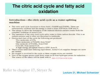
Links between the urea cycle and citric acid cycle. • The interconnection between urea cycle and citric acid cycle—in a process called “Krebs bicycle”. • Each cycle can operate independently and communication between them depends on the transport of key intermediates between the mitochondrion and cytosol.
Several enzymes of the citric acid cycle, including fumaraseand malate dehydrogenase, are also present as isozymes in the cytosol.
The Fumarate generated in cytosolic arginine synthesis can therefore be converted to malate in the cytosol, and these intermediates can be further metabolized in the cytosol or transported into mitochondria for use in the citric acid cycle.
Aspartateformed in mitochondria by transamination between oxaloacetate and glutamatecan be transported to the cytosol, where it serves as nitrogen donor in the urea cycle reaction catalyzed by argininosuccinate synthetase.
These reactions, making up the aspartate-argininosuccinate shunt, provide metabolic links between the separate pathways by which the amino groups and carbon skeletons of amino acids are processed.
The activity of the urea cycle is regulated at two levels • The flux of nitrogen through the urea cycle in an individual animal varies with diet. • When the dietary intake is primarily protein, the carbon skeletons of amino acids are used for fuel, producing much urea from the excess amino groups.
During prolonged starvation, when breakdown of muscle protein begins to supply much of the organism’s metabolic energy, urea production also increases substantially.