Download
1 / 26
310 likes | 964 Vues
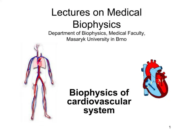
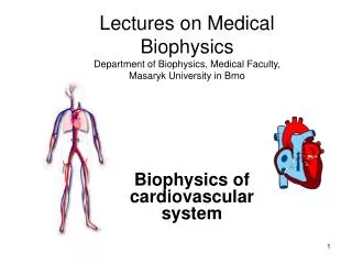
Lectures on Medical Biophysics Department of Biophysics, Medical Faculty, Masaryk University in Brno. Biophysics of cardiovascular system. Lecture outline. Mechanical properties of blood vessels Reynolds number Blood flow in blood vessels Peripheral resistance of blood vessels

E N D
Lectures on Medical BiophysicsDepartment of Biophysics, Medical Faculty, Masaryk University in Brno Biophysics of cardiovascular system
Lecture outline • Mechanical properties of blood vessels • Reynolds number • Blood flow in blood vessels • Peripheral resistance of blood vessels • Mechanical work and power of heart • Capillary ultrafiltration • Kidneys: renal work and glomerular ultrafiltration • Blood pressure measurement
Mechanical properties of cardiovascular system Closed circulation and transport system • Main parts: • Heart muscle (myocardium) • Closed system of blood vessels • Blood • Main functions: • Supplying cells by nutrients and oxygen, • Transport of hormones and other chemical signals, • Removal of waste and side products from cells (tissues) • Heat transfer - thermoregulation
Mechanical properties of blood vessels Tension T in walls of some blood vessels: • Laplace law– mechanical stress of blood vessel walls is directly proportional to the pressure and vessel radius
Elastic and muscular blood vessels Aorta behaves like typical elastic vessel Arterioles are muscular vessels able of vasodilatation and vasoconstriction
Reynolds number • Blood flow: laminar (ordered) • turbulent (whirling) • Reynolds (1883) • Reynolds number (for a liquid flowing in a cylindrical tube): Re = r.vm.r/h r – liquid density, vm – mean flow rate, r – vessel radius, h – coefficient of dynamic viscosity • Critical flow rate: vm = 1000h/r.r
Blood flow in blood vessels The differences between theoretical and real flow rate profiles are given mainly by the fact that blood is a non-Newtonian liquidand is influenced by the distensibility and compliance of the vessel wall.. The flow rate profile changes during pulse wave. We can obtain important diagnostic information from values of blood velocity and the shape of the flow rate curve.
Blood flow in an obstructed blood vessel Fig. after Cameron et al., 1999 The upper curve represents blood flow in a vessel without atherosclerotic stenosis (narrowing), the lower one in a vessel with stenosis. We need bigger increase of pressure Dp for the same increase in blood flow DQ (volume per time unit).
Peripheral impedance (resistance) of blood vessels • Analogy of electrical impedance (R = U/I) • Pressure p is an analogy of voltage U • Blood flow volume Q is an analogy of electric current I • R = Dp/Q • Considering the Hagen-Poiseuille formula for flow volume (m3.s-1):
Peripheral resistance of blood vessels • Low vascular impedance characteristic for brain, liver, spleen and kidney arteries • High vascular impedance characteristic for arteries of skeletal muscles
Peripheral resistance of blood vessels • Percentage of total peripheral resistance estimated for individual segments of blood circulation: • arteries ......... 66 % • (among those arterioles 40 %) • capillaries ........ 27 % • veins ............. 7 % • In vasodilatation, R decreases – heart load decreases • In vasoconstriction,R increases – heart load increases
Mechanical work and power of heart • Mechanical power of heart (for pulse rate 70 min-1) ........ 1,3 W • Total power of heart (at rest conditions) ......................13 W • Total power of human organism (at rest conditions) ............................................115 W • Mechanical work of heart muscle: Some very small work dW is done agains external pressure p during ejection of very small blood volume dV: dW = p.dV The whole work during a systole: W = p.dV Very small part of this work is transformed into kinetic energy of blood ejected.
Estimation of heart work during one systole • p = const. W = pm.DV, pm is mean blood pressure at the beginning of aorta • Left ventricle Right ventricle pm = 13.3 kPa pm. = 2.7 kPa DV = 70 ml DV = 70 ml W = 0.93 J W = 0,19 J • Mechanical energy of ejected blood volume Wk : = 0.009 J = 0,0018 J (since Wk = 1/2.r.vm2.DV, r = 1.06x10³ kg.m-3, vm. = 0.3 m.s-1, resp. 0.22 m.s-1 in pulmonary artery)
Work and efficiency of myocardium • Energy necessary to maintain tonus of myocardium: aT.dt • T – mechanical tension of heart walls (tonus) [N.m-1], t - time • Total energy necessary: Ec = p.dV + aT.dt • Mechanical efficiency: W/Ec(max. 10 %)
Filtration process in capillary loop Hydrostatic pressure: = 4.7 kPa = 2.3 kPa Oncotic pressure = 3.5 kPa filtration pressure capillary
!!!!!!!!!!!!! Oedemas arise due to low protein level in blood plasma, which causes low oncotic pressure and increases so the filtration pressure.
Kidneys: renal work and glomerular ultrafiltration • The osmotic work necessary to transfer a substance from a medium with substance concentration C2to the medium with substance concentration C1. (It is transfer of needful substances from primary urine back to the blood.) W = 2.3 nRT logC1/C2 • Glomerular ultrafiltration: Hydrostatic pressure in glomerularcapillaries is about 6.6 kPa (50 mmHg). The following pressures have opposite effect: hydrostatic pressurein Bowman’s capsule – 1.3 kPa (10 mm Hg) and oncotic pressure of plasma proteins – 3.3 kPa (25 mm Hg), so the resulting filtration pressure in glomerulus is 2 kPa (15 mmHg)under normal circumstances.
Glomerulushttp://coe.fgcu.edu/faculty/greenep/kidney/Glomerulus.htmlGlomerulushttp://coe.fgcu.edu/faculty/greenep/kidney/Glomerulus.html -1.3 kPa +2.0 kPa - 3.3 kPa +6.6 kPa
Blood pressure measurement • Pressure is defined as a force acting on unit area in a gas or liquid. p = F/A [N.m-2], where F is the force acting on the area A. • In the SI system, the pressure is measured in N.m-2, the unit is called pascal [Pa]. • The most common way of indicating pressure in medicine is by the height of mercury column in millimetres - mmHg. • 1 mmHg = 1 torr = 133.3 Pa
Blood pressure measurement • In arteries, the blood pressure oscillates between a maximum value, i.e. systolic pressure, and a minimum value of the pressure, i.e. diastolic blood pressure. • The time-course of the blood pressure changes is periodical but non-sinusiodal. • The difference between the systolic and diastolic pressures is maximal at the beginning of aorta; the pressure fluctuates in the range of 10.5 to 16 kPa, i.e. 80 to 120 mmHg. • The mean value of blood pressure in lung artery represents only one fifth of the blood pressure in the aorta.
Riva-Rocci method An inflatable cuff with manometer is put on the arm above the elbow (heart level), inflated to a pressure higher than the systolic pressure in a. brachialis. Blood flow is stopped. The pressure in the cuff is gradually decreased. At systolic pressure, blood starts to flow through the artery. The turbulent flow produces acoustic noise - Korotkoff sounds, audible in a stethoscope placed in the elbow pit. As pressure in the cuff is decreased the sounds become louder, culminate and gradually decrease, disappearing altogether at the diastolic pressure (laminar flow renewed). The maximum loudness is at mean arterial pressure.
Riva-Rocci method • The Riva-Rocci method can be objectified and automatised for patient monitoring. The cuff is regularly inflated by a small compressor (e.g., every ten minutes) and the Korotkoff sounds are recorded by a microphone. Measured values of systolic and diastolic pressures can be displayed (in simple devices) or stored in the instrument memory and evaluated later. This method is called Holter monitoring of blood pressure. • In small children, this auscultation method can fail. In this case, we can use the Doppler flow detectors to detect blood flow in the artery compressed by the cuff.
Direct measurement of blood pressure • Invasive measurement of BP is a direct method. A thin, flexible catheter or probe must be inserted into the blood vessel. Its free end usually is connected to a transducer (capacity or piezoelectric) but it is also possible to insert a piezoelectric transducer directly into the vessel. This method is rather risky, so that it is used relatively seldom. However, it is the only method which allows measurements of pressure in veins or inside the heart.
Author: Vojtěch MornsteinContent collaboration C.J. Caruana, I. Hrazdira,language revision: C.J. CaruanaPresentation design: - - -Last revision: January 2012