Download
1 / 21
270 likes | 640 Vues
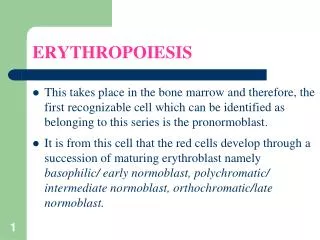
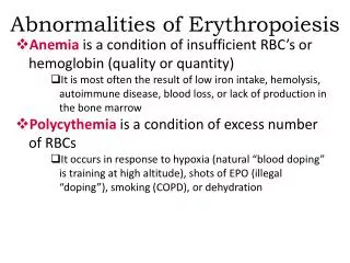
Abnormalities of Erythropoiesis. Anemia is a condition of insufficient RBC’s or hemoglobin (quality or quantity) It is most often the result of low iron intake, hemolysis, autoimmune disease, blood loss, or lack of production in the bone marrow

E N D
Abnormalities of Erythropoiesis • Anemia is a condition of insufficient RBC’s or hemoglobin (quality or quantity) • It is most often the result of low iron intake, hemolysis, autoimmune disease, blood loss, or lack of production in the bone marrow • Polycythemiais a condition of excess number of RBCs • It occurs in response to hypoxia (natural “blood doping” is training at high altitude), shots of EPO (illegal “doping”), smoking (COPD), or dehydration
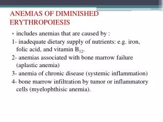
Anemias • Iron deficiency anemia is the most common anemia in the U.S., and affects primarily menstruating women • In the United States, 20% of all women of childbearing age have iron deficiency anemia, compared with only 2% of adult men • Hemorrhagic anemia is the result of precipitous blood loss, and results in an equal decrease in Hct, Hgb content, and RBC count
Anemias • Sickle-cell disease (SCD), also called sickle-cell anemia, is an autosomal recessive disorder. A genetic defect in the primary DNA sequence leads to production of a faulty Hgb β chain, and RBCs that take on a rigid, sickle-shape • Sickling decreases the cells' flexibility and results in a variety of complications; life expectancy is shortened
RBC Life Cycle • RBCs live only about 120 days. To maintain normal numbers, new mature cells must enter the circulation at the astonishing rate of at least 2 million/second, a pace that balances the equally high rate of RBC destruction • Ruptured RBCs are removed from circulation and destroyed by fixed phagocytic macrophages in the spleen and liver—the breakdown products are recycled and used in numerous metabolic processes, including the formation of new RBCs
RBC Life Cycle Circulation for about 120 days Circulation for about 120 days Circulation for about 120 days Circulation for about 120 days Circulation for about 120 days Circulation for about 120 days 7 7 7 7 7 7 7 3 3 3 3 3 3 3 3 3 3 3 Reused for protein synthesis Reused for protein synthesis Reused for protein synthesis Reused for protein synthesis Reused for protein synthesis Reused for protein synthesis Reused for protein synthesis Reused for protein synthesis Reused for protein synthesis Reused for protein synthesis Reused for protein synthesis Amino acids Amino acids Amino acids Amino acids Amino acids Amino acids Amino acids Amino acids Amino acids Amino acids Amino acids Transferrin Transferrin Transferrin Transferrin Transferrin Transferrin Transferrin Transferrin Fe3+ Fe3+ Fe3+ Fe3+ Fe3+ Fe3+ Fe3+ Fe3+ Globin Globin Globin Globin Globin Globin Globin Globin Globin Globin Globin Globin 6 6 6 6 6 6 6 6 4 4 4 4 4 4 4 4 4 4 5 5 5 5 5 5 5 5 5 Fe3+ Fe3+ Fe3+ Fe3+ Fe3+ Fe3+ Fe3+ Fe3+ Fe3+ Fe3+ 2 2 2 2 2 2 2 2 2 2 2 2 Fe3+ Fe3+ Fe3+ Fe3+ Fe3+ Fe3+ Fe3+ Ferritin Ferritin Ferritin Ferritin Ferritin Ferritin Ferritin Ferritin Ferritin Heme Heme Heme Heme Heme Heme Heme Heme Heme Heme Heme Heme Transferrin Transferrin Transferrin Transferrin Transferrin Transferrin Transferrin Transferrin Transferrin Transferrin + + + + + + + Globin Globin Globin Globin Globin Globin Globin Bilirubin Bilirubin Bilirubin Bilirubin 9 9 9 9 9 + + + + + + + Biliverdin Biliverdin Biliverdin Biliverdin Biliverdin Liver Liver Liver Liver Liver Liver Liver Liver Liver Vitamin B12 Vitamin B12 Vitamin B12 Vitamin B12 Vitamin B12 Vitamin B12 Vitamin B12 Bilirubin Bilirubin Bilirubin Bilirubin Bilirubin 11 11 11 1 1 1 1 1 1 1 1 1 1 1 1 1 Red blood cell death and phagocytosis Red blood cell death and phagocytosis Red blood cell death and phagocytosis Red blood cell death and phagocytosis Red blood cell death and phagocytosis Red blood cell death and phagocytosis Red blood cell death and phagocytosis Red blood cell death and phagocytosis Red blood cell death and phagocytosis Red blood cell death and phagocytosis Red blood cell death and phagocytosis Red blood cell death and phagocytosis Red blood cell death and phagocytosis + + + + + + + 10 10 10 10 Erythopoietin Erythopoietin Erythopoietin Erythopoietin Erythopoietin Erythopoietin Erythopoietin Small intestine Small intestine Small intestine Kidney Kidney 8 8 8 8 8 8 Erythropoiesis in red bone marrow Erythropoiesis in red bone marrow Erythropoiesis in red bone marrow Erythropoiesis in red bone marrow Erythropoiesis in red bone marrow Erythropoiesis in red bone marrow Bilirubin Bilirubin Bilirubin 13 13 12 12 12 Urobilin Urobilin Urobilinogen Urobilinogen Urobilinogen Macrophage in spleen, liver, or red bone marrow Macrophage in spleen, liver, or red bone marrow Macrophage in spleen, liver, or red bone marrow Macrophage in spleen, liver, or red bone marrow Macrophage in spleen, liver, or red bone marrow Macrophage in spleen, liver, or red bone marrow Macrophage in spleen, liver, or red bone marrow Macrophage in spleen, liver, or red bone marrow Macrophage in spleen, liver, or red bone marrow Macrophage in spleen, liver, or red bone marrow Macrophage in spleen, liver, or red bone marrow Macrophage in spleen, liver, or red bone marrow Macrophage in spleen, liver, or red bone marrow Key: Key: Key: Key: Key: Key: Key: Key: Key: Key: Key: Key: Key: Bacteria Bacteria Bacteria in blood in blood in blood in blood in blood in blood in blood in blood in blood in blood in blood in blood in blood Stercobilin Stercobilin Stercobilin 14 Large intestine in bile in bile in bile in bile in bile in bile in bile in bile in bile in bile in bile in bile in bile Feces Feces Feces Urine Urine
Leukocytes • Unlike RBCs, white blood cells (WBCs)or leukocyteshave nuclei and a full complement of other organelles - but they do not contain the protein Hgb
Leukocytes • Leukocytes are divided into two groups depending on whether they contain conspicuous chemical-filled cytoplasmic granules (when stained) • Granulocytes include the neutrophils, eosinophils, and basophils • Agranulocytes are the monocytes and lymphocytes
Leukocytes • The most numerous WBC in normal blood (60-70% of circulating white cells) is the neutrophil, or polymorphonucleocyte (PMN) • PMNs are granulocytes with a pinkish cytoplasm, and they are one of the two major phagocytes in the body • their principal role is to fight bacterial infections
Leukocytes • Chemicals released by microbes and inflamed tissues attract phagocytes, a phenomenon called chemotaxis • This graphic shows a PMN phagocytizing a microbe for internal digestion and destruction
Leukocytes • Eosinophils are characterized by their large red granules • They are much less numerous than neutrophils (2-4% of circulating WBCs), but their numbers increase slightly with parasitic infection • they have also been associated with the development of allergies
Leukocytes • While monocytesare not granulocytes, they come from the same immediate precursor cell as the 3 granulocytes (the myeloid stem cell) • Along with neutrophils, monocytes are the other major group of phagocytic cells. Even though they constitute only 3-8% of the circulating WBCs, they are much more numerous in the peripheral, tissues where they act as “fixed” phagocytes
Leukocytes • Lymphocytes are the last of the 5 types of WBCs, and in many ways they are quite different • Lymphocytes don’t have granules or phagocytize; their cytoplasm is sparse compared to their very large nucleus, and they develop from a different precursor stem cell • Also, rather than acting as non- specific defenders, lymphocytes develop as responders to very specific foreign antigens
Leukocytes • Basophils are the third type of granulocyte; they contain large, dark blue, histamine containing granules • Normally, they are the lowest number of circulating WBCs (only 0-1%), but they have an important role to play in the inflammatory responses
Leukocytes • Approximately 20-30% of circulating white cells are lymphocytes: an increase above this number is called a lymphocytosis and often represents an acute viral infection • Most lymphocytes continually move among lymphoid tissues, lymph, and blood, spending only a few hours at a time in blood • Lymphocytes are the cornerstone of the specific immune response
WBC Indices • For diagnostic purposes, physicians measure the total number of circulating WBCs • A leukocytosisis any WBC count > 10,000/mm3, and usually indicate an infectious process or a cancer • A leukopenia is any WBC count < 5,000/mm3, and usually indicates a severe disease (AIDS, bone marrow failure, severe malnutrition, or chemotherapy)
WBC Indices • To enhance the diagnostic value of a WBC count, the percentages of each of the 5 types of WBCs is determined by using a machine to do a statistical analysis of the blood sample. This is called the WBC differential
WBC Indices • Shifts in the normal percentages of circulating WBCs will often point towards a bacterial infection (elevated percentage of neutrophils) or a viral infection (elevated percentage of lymphocytes • In this peripheral blood smear a patient with lymphocytic leukemia has a WBC >150,000 and 90% of the WBCs are cancerous lymphocytes! Lymphocytic leukemia.
Plasma • Plasma is the fluid component of the blood and contains everything in blood except the formed elements, which, for collection purposes, have been centrifuged out • Plasma contains mostly water, with electrolytes, hormones, proteins, dissolved gasses, and glucose and other nutrients
Plasma Proteins • The major protein in plasma is albumin; it also has many clotting proteins, antibodies, and enzymes • Albumin is a relatively simple, water soluble protein with a low molecular weight – it forms small heart-shaped globules just over 8 nm in size • Albumin is synthesized in the liver and contributes significantly to the blood viscosity and the body’s ability to maintain blood pressure • It also plays an important role as a carrier molecule
Plasma Proteins • Another important group of plasma proteins are the globulins, of which there are several types: α (alpha), β (beta), and δ (gamma). Globulins control blood osmotic pressure and act as carrier molecules • α-globulins carry bilirubin and steroids • β-globulins carry copper and iron • δ-globulins are immunoglobulins (antibodies) made by activated B lymphocytes called plasma cells
Hemostasis • Hemostasis is a sequence of responses that stops bleeding • When blood vessels are damaged or ruptured, the hemostatic response must be quick, localized to the region of damage, and carefully controlled in order to be effective • Three mechanisms reduce blood loss • Vascular spasm • Formation of a platelet plug • Blood clotting (coagulation)