Upper Gastrointestinal Emergencies
Upper Gastrointestinal Emergencies. Author: Andrew McDonald MD, FRCP, Assistant Professor Date Created: January 2012. Learning Objectives. Understand the approach to GI bleeding Understand the approach to esophageal injuries from caustics and foreign bodies

Upper Gastrointestinal Emergencies
E N D
Presentation Transcript
Upper Gastrointestinal Emergencies Author: Andrew McDonald MD, FRCP, Assistant Professor Date Created: January 2012
Learning Objectives • Understand the approach to GI bleeding • Understand the approach to esophageal injuries from caustics and foreign bodies • Understand the approach to peptic ulcer disease and gastritis
Case example • A 31 year old man is brought by his family after vomiting black material for two days • He appears unwell and lethargic • HR 130 BP 90/50 RR 30 T 35°C • Family says he has a history of chronic liver disease
GI bleeding – How patients present • History of vomiting blood or rectal blood • Shock +/- passing blood • Decreased LOC +/- passing blood
Challenges in these patients • Management of hypovolemic shock • Vomiting and aspiration • Hepatic encephalopathy • Coagulation disorder
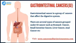
Causes of Upper GI bleeding • Peptic ulcer disease • Gastritis • Varices • Mallory – Weiss tear rare • Malignancies
Causes of Lower GI bleeding • Hemorrhoids • Diverticulosis • Malignancies/polyps • Angiodysplasia (AVM) of aging • Inflammatory bowel disease • Complications of Typhoid fever • Upper GI bleeding • Bloody diarrhea
Epidemiology • Little is documented on the epidemiology of GI bleeding in developing countries
Clinical features • Hematemesis = upper GI source • Hematochezia = lower GI source • Melena = don’t know source
Clinical features (continued) • Weight loss -- Think of malignancy • Bleeding following vomiting -- Think of Mallory Weiss tear • Medications can cause bleeding: • NSAID/ASA • Steroids • Anticoagulants • Alcohol use/abuse associated with various types of bleeding
Clinical features (continued) • Establish vascular volume status • Confirm bleeding by site • Do a rectal exam to look for bright red blood or melena; perform a guaiac test if available • Role for NG tube? • Look for signs of liver disease • Look for generalized bleeding problem
Management • Assess for airway management • Prompt large bore iv access • Volume resuscitation if necessary as patients can deteriorate rapidly • CBC, cross match, LFT, coagulation, renal • Reverse any coagulopthy if possible • Access to endoscopy as diagnostic and therapeutic procedure (Ideal <24 hours)
Management (cont.) - Medications • Reducing gastric acidity via H2 blockers or PPI meds • Reducing portal pressure for varices • Antibiotics may improve survival • Use of Sengstaken-Blakemore tube not recommended due to complications • Need for surgery uncommon
Case continued • Patient’s airway reflexes were intact • Given Oxygen for shock state • Monitored vascular/respiratory status closely • Administered fluids to improve perfusion • Cross matched for blood and plasma to restore hemoglobin and coagulation • PPI and antibiotics given while waiting for endoscopy
Esophageal emergencies • Causes: • Varices • Ingestion of corrosives • Foreign bodies
Caustics – how patients present • Pain • Difficulty swallowing • Airway compromise
Challenges in these patients • Protecting healthcare workers • Pain masking complications • Systemic effects of chemical/co-ingestion • Mental health issues
Causes • Intentional self harm versus accidental • Sources of chemical information
Causes (continued) • Alkali – liquefaction necrosis, thrombosis • Acids – coagulation necrosis, eschar, systemic absorption
Clinical features • Pain – range of severity • Respiratory/airway symptoms • GI symptoms Absence of oral injury does not preclude GI injury!
Management • Protect yourself • Airway assessment – direct vision technique • Treat shock = GI bleed, perforation, delayed sepsis, metabolic • Decontaminate eyes and skin as needed • Surgical consult if perforation
Esophageal FB – How patients present • Usually based on history • Chest pain, retching, can’t swallow • Beware of children, mental health, “prisoners”
Clinical features • Problems with handling secretions • Location in esophagus • Pediatric typically proximal • Adults typically distal • Perforation is uncommon • Endoscopy is diagnostic and therapeutic procedure
Diagnosis • X-ray can show the location of a foreign body
Management • Endoscopy preferred • Time +/- sedation often works • Meds: • Glucagon 1 mg IV • Nifedipine 10 mg SL • Nitroglycerine SL
Management (continued) • Button batteries and coins: • Remove if in esophagus if endoscopy available • Remove if still in stomach after 24 h • Sharp objects • Endoscopy preferred if available
Ulcers and gastritis – How patients present • Pain • GI bleeding • Perforation (shock)
Causes • H. pylori infection • Meds: • NSAID/ASA • Alcohol • Spices • Severe physiological stress
Clinical features • Pain • Often epigastric tenderness without peritonitis • Tests not really useful except to rule out other things
Management • Perforation, bleeding discussed elsewhere • Antacids • H2 blockers, PPI • Antibiotic therapy • Avoidance of NSAID and alcohol
Quiz Question 1 • Which is the most common cause of upper GI bleeding? • Malignancy • Intestinal perforation • Peptic ulcers/gastritis • Mallory Weis tear
Quiz Question 2 • GI bleeding can present as: • Melena • Hematemesis • Shock without obvious blood loss • Hematochezia • All of the above are correct
Quiz Question 3 • In managing patient after a caustic ingestion: • They usually present with shock • Those without any pain are the sickest • Their vomit can be harmful to care givers • An NG tube should always be placed
Quiz Question 4 • Regarding esophageal obstruction: • Endoscopy is never indicated • If batteries are notobstructing the esophagus, they can be left there for up to three days • Adults and children usually obstruct proximally • All patients with obstruction should be intubated • Medications may sometimes prevent the need for endoscopy
Quiz Question 5 • Regarding patients with peptic ulcer disease: • Abdominal pain is usually constant • Alcohol use is one of the causes of ulcers • Acetaminophen is a common cause of ulcers • The usual treatment is surgical repair
Summary • GI bleeding can be a cause of life-threatening shock requiring resuscitation • Esophageal injuries should be managed in conjunction with endoscopy experts • Peptic ulcer disease and gastritis can present as life-threatening complications
General References • Tintinalli, JE et al (2011) Chapters 78, 79, 80, 81, 194. McGraw Hill Publishers Emergency Medicine – A study guide 7th Edition, USA • Manson’s Tropical Diseases, Chapter 10. Saunders Elsevier, 22nd edition.