Download

1 / 33
330 likes | 361 Vues
Learn about Respiratory Acidosis, the imbalance in CO2 production and excretion in the body leading to low blood pH. Understand the regulation and control mechanisms, types of disorders, and associated diseases. Discover how respiratory diseases impact ventilation and gas exchange. Get insights into causes and management strategies.

E N D
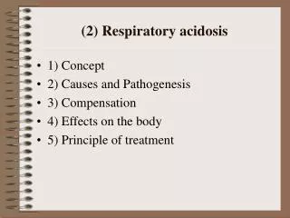
RESPIRATORY ACIDOSISLt Col (R) Dr. Anjum IqbalAssociate Professor PathologyWah Medical College
RESPIRATORY ACIDOSIS Respiratory acidosis occurs when the arterial partial pressure of carbon dioxide (PaCO2) is elevated above the normal range (>45 mm Hg) leading to a blood pH less than 7.35.
RESPIRATORY ACIDOSIS Respiratory acidosis is not a specific disease. Instead, it is an abnormality resulting from an imbalance between carbon dioxide (CO2) production by the body and excretion by the lungs.
REGULATION OF ATERIAL PCO2 • Rate of CO2 production closely parallels energy requirement. • It diffuses out of cells, into the interstitial fluid and then to capillaries. • Blood entering the tissue capillaries PCO2 of about 40 mm Hg. • Diffusion of CO2 into capillaries raises PCO2 to 45 mm Hg.
REGULATION OF ATERIAL PCO2 • Alveolar air has PCO2 of about 40 mm Hg • 5 mm Hg diffusion gradient. • Pulmonary vein PCO2 is about 40 mm Hg.
VENTILATORY RATE & PCO2 • To hold the PCO2 of blood constant, the body must keep the rate of CO2 production & excretion closely matched. • Achieved by changing the rate of alveolar ventilation. • Arterial PCO2 responds inversely to ventilatory rate: a high rate of ventilation yields low PCO2.
PHYSIOLOGIC CONTROL • Matching ventilatory rate to CO2 production is achieved by feedback from: • PCO2 – sensitive chemoreceptors. • The carotid bodies. • The chemoreceptors innervate respiratory centres in the brain stem, which control the rate and depth of breathing.
FUNDAMENTAL CONCEPT PCO2 CO2 Production / Alveolar ventilation • If CO2 production rises and alveolar ventilation is constant, arterial PCO2 rises. (Acidosis) • If CO2 production is constant and alveolar ventilation falls, PCO2 rises. (Acidosis) • If CO2 production is constant and alveolar ventilation rises, PCO2 falls. (Alkalosis) • Alveolar ventilation is responsible for CO2 elimination and is calculated when the respiratory frequency is multiplied by the difference between the tidal volume and the physiologic dead space.
NATURE OF RESPIRATORY DISEASE • There are three types of problems: 1. Neuromuscular chain defects. 2. Pulmonary diseases 3. Increased CO2 production.
Neuromuscular chain defects • Normal ventilation requires an intact neuromuscular chain. • When a link in the neuromuscular chain fails, the minute ventilation decrease. • This decrease may occur via a fall in either tidal volume or respiratory rate or both.
Neuromuscular chain defects • When minute ventilation decreases, alveolar ventilation decreases with it. • The fall in alveolar ventilation causes PCO2 to rise, resulting in hypercapnia • If the function of any part of the chain is impaired, even slightly, it may be impossible to raise minute ventilation enough to match a rising ventilatory requirement.
Pulmonary diseases • Pulmonary diseases refers specifically to defects in the lung itself and typically affect the lung with uneven severity, so some areas are damaged and others are relatively normal. • The five major types of defects are: 1. Air way obstruction. 2. Alveolar filling. 3. Atelectasis. 4. Perfusion blockage 5. Loss of lung parenchyma
Air way obstruction • There is narrowing of the airway that supplies one region of alveoli. • Narrowing may be due to blockage in the lumen, thickening of the air way wall, or from contraction of smooth muscle in the airway walls. • It reduces the amount of fresh air that reaches the alveoli, causing “localized hypoventilation”. • Examples of diseases include asthma and chronic bronchitis.
Alveolar filling • Alveoli may be filled with liquid or semi liquid material. • Air is entirely prevented from reaching the alveolar surface. • The blood vessels act as “shunt” • It is an extreme form of localized hypoventilation. • Examples: Severe pulmonary edema and severe pneumonia
Atelectasis • There may be collapse of air spaces. • If collapse is partial, ventilation is reduced. • If collapse is total, ventilation is eliminated and area acts like a shunt. • Example: Collapsed lung.
Perfusion blockage • In perfusion (blood flow) blockage, ventilation is normal but the loss of blood flow makes the alveoli completely ineffective as a gas exchange surface. • Perfusion blockage turns alveoli into dead space. • Example: Pulmonary thromboembolism.
Loss of lung parenchyma • When there is loss of alveolar walls and capillaries, this wall loss greatly reduces the area of the lung’s exchange surface, creating abnormally large air spaces in the lung (dead space). • Example: Emphysema.
Increased CO2 production • Common causes of increased CO2 production include: - Fever - Physical activity - Hyperthyroidism - Overfeeding with too many calories.
RESPIRATORY ACIDOSIS • In heath, strength is well matched to load. • In respiratory diseases, load may exceed strength. • The neuromuscular chain cannot maintain output at a level that meets the increased ventilatory requirement.
RESPIRATORY ACIDOSIS • Basic cause of Respiratory acidosis is: - Increase in PaCO2 • Which results in: - Decrease in pH - Slight increase in HCO3- • Normal Systemic Arterial Values: - PCO2 - 40 mm of Hg (35 - 45) - HCO3- - 24 mmol/l (21 - 29)
TYPES OF RESPIRATORY ACIDOSIS • Respiratory acidosis can be: - Acute Respiratory acidosis - Chronic Respiratory acidosis
ACUTE RESPIRATORY ACIDOSIS In acute respiratory acidosis, the PaCO2 is elevated above the upper limit of the reference range (ie, >45 mm Hg) with an accompanying acidemia (ie, pH <7.35).
ACUTE RESPIRATORY ACIDOSIS • Acute respiratory acidosis occurs when an abrupt failure of ventilation occurs. • This failure in ventilation may be caused by : - Depression of the central respiratory center by cerebral disease or drugs - Inability to ventilate adequately due to neuromuscular disease (e.g, myasthenia gravis, amyotrophic lateral sclerosis, Guillain-Barré syndrome, muscular dystrophy). - Airway obstruction related to asthma or chronic obstructive pulmonary disease (COPD) exacerbation.
ACUTE RESPIRATORY ACIDOSIS In acute respiratory acidosis, compensation occurs in 2 steps. • The initial response is cellular buffering that occurs over minutes to hours. • Cellular buffering elevates plasma bicarbonate (HCO3-) only slightly, approximately 1mEq/L for each 10-mm Hg increase in PaCO2.
ACUTE RESPIRATORY ACIDOSIS • The second step is renal compensation that occurs over 3-5 days. • With renal compensation, renal excretion of carbonic acid is increased and bicarbonate reabsorption is increased. • In renal compensation, plasma bicarbonate rises 3.5 mEq/L for each increase of 10 mm Hg in PaCO2.
ACUTE RESPIRATORY ACIDOSIS • The expected change in serum bicarbonate conc. in acute respiratory acidosis can be estimated as follows: - HCO3- increases 1 Eq/L for each 10-mm Hg rise in PaCO2. • The expected change in pH with acute respiratory acidosis can be estimated with the following equations: • Change in pH = 0.008 X (40 – PaCO2)
CHRONIC RESPIRATORY ACIDOSIS In chronic respiratory acidosis, the PaCO2 is elevated above the upper limit of the reference range, with a normal or near-normal pH secondary to renal compensation and an elevated serum bicarbonate (ie, HCO3- >30 mm Hg).
Chronic respiratory acidosis • Chronic respiratory acidosis may be secondary to many disorders, including : - - COPD. - Obesity hypoventilation syndrome (ie, pickwickian syndrome) - Neuromuscular disorders such as amyotrophic lateral sclerosis - severe restrictive ventilatory defects as observed in interstitial fibrosis and thoracic deformities.
Chronic respiratory acidosis • The expected change in serum bicarbonate conc. in chronic respiratory acidosis can be estimated as follows: - HCO3- rises 3.5 mEq/L for each 10 mm Hg rise in PaCO2. • The expected change in pH with chronic respiratory acidosis can be estimated with the following equations: - Change in pH = 0.003 X (40 - PaCO2)
EFFECTS OF ACIDOSIS ON ELECTROLYTES • Respiratory acidosis does not have a great effect on electrolyte levels. • Some small effects occur on calcium and potassium levels. • Acidosis decreases binding of calcium to albumin and tends to increase serum ionized calcium levels. • Acidemia causes an extracellular shift of potassium, but respiratory acidosis rarely causes clinically significant hyperkalemia.
RESPIRATORY ACIDOSIS N = Normal values A = Uncompensated respiratory acidosis (Acute Hypoventilation) B = Partially Compensated respiratory acidosis. C = Completely Compensated respiratory acidosis.