Download
1 / 73
860 likes | 1.38k Vues
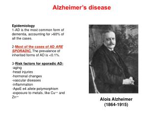
Alzheimer’s disease. Epidemiology 1-AD is the most common form of dementia, accounting for >60% of all the cases. 2- Most of the cases of AD ARE SPORADIC . The prevalence of inherited forms of AD is <0.1%. 3- Risk factors for sporadic AD: -aging -head injuries -hormonal changes

E N D
Alzheimer’s disease Epidemiology 1-AD is the most common form of dementia, accounting for >60% of all the cases. 2-Most of the cases of AD ARE SPORADIC. The prevalence of inherited forms of AD is <0.1%. 3-Risk factors for sporadic AD: -aging -head injuries -hormonal changes -vascular diseases -inflammation -ApoE e4 allele polymorphism -exposure to metals, like Cu++ and Zn++ Alois Alzheimer (1864-1915)
First description of tangle and plaque pathology by Alois Alzheimer (1901) A nissl stain tissue section (left) and a Bielkhowsky’s stain section from Augustine Deter’s brain (right) http://scienceblogs.com/neurophilosophy/Alzheimer
Alzheimer’s disease AD is a progressive neurodegenerative disorder affecting the elderly population. Once it starts, it progresses with aging. It is characterized by the presence of lesions both at an extracellular level (the b-amyloid plaques), and at an intracellular levels (the Neurofibrillary tangles, NFT). The presence of the lesions correlates with both reduction in the volume of the brain (as a consequence of the neurodegenerative phenomena), and with cognitive decline associated to loss of memory. AD is a form of Dementia.
Definition of Dementia The loss of intellectual functions (such as thinking, remembering, and reasoning) of sufficient severity to interfere with a person’s daily functioning. Dementia is not a disease itself but rather a group of symptoms that may accompany certain diseases or conditions. Symptoms may also include changes in personality, mood, and behavior. Dementia is irreversible when caused by disease or injury but may be reversible when caused by drugs, alcohol, hormone or vitamin imbalances, or depression.
Symptoms in AD Increasing and persistent forgetfulness Difficulty performing familiar tasks Problems with finding the right words to express your thoughts Disorientation with time and place Poor or impaired judgment Problems with abstract thinking Putting everyday items in illogical places Mood, behavior or personality changes.
Comparison between AD signs and age-related memory changes
Anatomy of the Alzheimer’s disease brain PET MRI Normal AD www.elements4health.com
Deposition of fibrillar proteinacious material in Alzheimer’s disease Alzheimer’s disease: characterized by extracellular depositions, the b-amyloid plaque, and intracellular depositions, the Neurofibrillary Tangles(NFT) comprised of Paired Helical Filaments (PHF), aggregates of hyperphosphorylated protein tau.
Origin of NFTs and the mechanism of tau pathology 1- Tau is a microtubule-associated protein that regulates cytoskeleton structure. 2- Tau is hyperphosphorylated in AD. 3- When highly phosphorylated, tau is sequestered into aggregates (PHF and NFT) and causes disruption of microtubules, that ultimately leads to cell death. www.emdbiosciences.com
Origin of the b-amyloid plaque 1-Amyloidogenic processing of the Amyloid Precursor Protein APP, and generation of the the b-amyloid peptides (Ab37-43). 1-bsecretase 2-gsecretase APP TMD The b-Amyloid Plaque NH2 COOH Ab amyloidogenic processing pathway Mechanisms of Ab pathology: Plaque: unknown Ab: oligomers affect synaptic activity and neurotransmitter release
The b-amyloid plaque 1- b-amyloid is DIFFERENT from amyloid. b-amyloid contains specifically the b-amyloid peptide, an approximately 4kDa peptide (Ab40, Ab42) deriving from the amyloid precursor protein APP when it undergoes the so called amyloidogenic pathway. b-amyloid plaque contains also ubiquitin and other proteins coming from degenerative neurons and glial cells. 2- The b-amyloid peptide is insoluble in water. When released from the precursor protein it assumes a b-sheet conformation that makes it hydrophobic. 3- b-amyloid peptides forms oligomers that may affect neurotransmitter release and synaptic plasticity. Oligomers will further aggregates in larger structures called fibrils, that will form the core of the b-amyloid plaque, depositing in the extracellular matrix. 4- The size of the plaque will increase following the progression of the disease. Scientific clear evidences are still missing in order to understand whether the plaque is a consequence or a cause of AD. Indeed, Ab andplaques formation are associated to neuronal death. Inherited forms of AD lead to substantial increase of Ab production.
Where do b-Amyloid peptides come from? From the processing so called AMYLOIDOGENIC of the larger precursor APP (Amyloid Precursor Protein)
Amyloidogenic processing of APP b-secretase g-secretase APP TMD b-secretase C99 NH2 COOH g-secretase Ab sbAPP COOH NH2
25-35, the most critical region for the change of conformation from a-sheet to b-sheet Structure of the b-Amyloid peptide
Genetic of AD -Depending on the kind of mutation and on the gene that carries it, the onset of the disease is SIGNIFICANTLY EARLIER than in sporadic cases (<before 65 years of age). All the mutations are autosomal dominant, and involve *APP protein *Members of the g-secretase complex, like Presenilin 1 (PS1) and Presenilin 2 (PS2)
Mutations on APP Chromosome 21. Relation to Down’s syndrome (Trisomy 21; excess of APP transcript induces plaque formation already at early age, and progresses with aging). Swedish mutation: K670M/N671L, facilitates the amyloidogenic processing of APP by increasing the affinity for and the cleavage by b-secretase of ~100 times. Dutch Mutation: E693Q, induces cerebral amyloidosis Arctic Mutation:E693G enhances b-Amyloid protofibril formation London Mutation: V717I, Affects the cleavage of g-secretase K670M/N671L V717I London Mutation E693G Arctic Mutation Swedish Mutation
Amyloidogenic processing of APP !!!b-secretase!!! !!!g-secretase!!! APP TMD b-secretase C99 NH2 COOH g-secretase Ab sbAPP COOH NH2 K670M/N671L V717I E693G
Mutations on Presenilins PS1 (on chromosome 14) PS2 (on chromosome 1) are ~450 aa long aspartyl proteases comprised of 7- to 8- transmembrane spanning domains. They are the catalytic part of a tetrameric protein complex called g-secretase, able to cleave APP at the end of the sequence for b-Amyloid peptides, generating b-Amyloid species. More than 70 mutations on PS1 gene account for inherited AD. Two mutations on PS2 gene account for inherited AD.
PS1 mutations and APP Hardy and Selkoe, 2002
Amyloidogenic processing of APP b-secretase g-secretase APP TMD NH2 COOH b-secretase C99 sbAPP COOH NH2 g-secretase/mutantPS1 Ab
The b-Amyloid Plaque Alois Alzheimer: “miliary bodies (the plaques) and dense bundles of fibrils (NFT)”
Characteristic of the b-amyloid plaque -is extracellular -normally, it has a core composed of the b-amyloid peptide -it can be cored (with an intensely stained core with a weak periphery stain-halo) or diffused, which do not have a core and the immunoreactivity is uniform over the plaque. -aggregation is massive in the center, and diffused at the sides (disaggregation hypothesis?) -diameter ranges between 20mm and 90mm -is composed of dystrophic neurites, b-amyloid peptides (40/42/43), ubiquitin, tau protein and other proteins, some involved in the generation of b-amyloid, like the secretases.
Amyloid: definition Insoluble fibrous protein aggregations sharing specific structural traits: -Insolubility in water -b-sheet conformation -largest aggregates deposit extracellularly -positive to the staining with specific dyes, such as Congo Red -associated with tissue degeneration (cause or consequence?) b-Amyloid Are formations of amyloid characterized by a core specifically formed by b-amyloid peptides, a product of the so called amyloidgenic processing of a larger precursor, the protein APP (Amyloid Precursor Protein)
Congo red staining of amyloid plaques Under polarized light Under unpolarized light
Congophylic plaques AD Prion’s disease Unpolarized light Polarized light
How to specifically detect b-Amyloid plaques? Using antibodies that specifically recognize the different b-Amyloid peptides, Ab 1-40, Ab 1-42, Ab 1-43
b-Amyloid peptides 40/42 43
Levels of deposited Ab40, Ab42 and Ab43 follow the progression of the disease
Diagnosis of AD At the moment, the exact diagnosis for AD can be performed only post-mortem by means of immunohistochemistry, verifying the presence of the b-amyloid plaques in the patient’s brain section using specific anti b-amyloid antibodies. However, a clinical diagnosis for AD is based on Neuropsycologic evaluation: behavioral and cognitive tests (MMSE, CDR). Diagnosis of a possible or probable AD (“Dementia of AD type”). Imaging techniques: MRI to detect changes in the volume of the brain PET analysis to identify areas of the brain with reduced glucose utilization Progress has been done in the direction of using dyes/compounds that specifically recognize the b-amyloid structure in both MRI and PET analysis. This would improve the quality of the diagnosis, identifying the structures (b-amyloid plaques) that are specific for AD.
Normal Alzheimer’s diseases MRI in Alzheimer’s disease (AD) MRI Computer graphic
Cerebral volume loss is associated with aging, but not necessarily with neuronal death and/or with AD. Need for diagnostic tools more specific than MRI.
Characteristics of a biomarker for diagnostic purposes -It should be already changed in the initial steps of the disease: implications for an early diagnosis of the disease, with deep impact in the success of the treatment. -The method to access the biomarker must not be invasive -The molecule used as a biomarker must be specific for that disease, and not be involved in other diseases. The mechanisms behind the specific changes in the disease must be clear. -The method of analysis must be reproducible :-) and must not be expensive -Ideally, the levels of the biomarker, once identified, should follow the progression of the disease (linear progression).
Possible biomarkers in AD • -Levels of b-amyloid peptides in the CSF of patients. Concerns regarding: • The method used to take the sample of CSF (spinal injection, which is invasive). • In the CSF of AD patients, the levels of the different species of b-amyloid peptides, in particular the most aggressive Ab42, do not always follow the progression of the disease. In fact, levels of Ab42 raise in the early stages of the diseases, but drop in the late, advanced stages of the disease, maybe indicating increased deposition of secreted Ab42 into plaques in the brain. • It is very difficult to correlate the amount of biomarker (in particular Ab42) to the amount of deposited plaques in the different stages of the disease. • Cerebrospinal levels of hyperphoshorylated tau. However, since tau is involved also in other neurodegenerative phenomena called taupaties, it is questionable how it could be considered as a biomarker specific for AD.
18 predictive AD-related plasma signaling molecules Hematopoiesis Apoptosis Immune response
Probable AD diagnosis via identification of changes in the 18 markers Ray et al.,
The Pittsburgh Compound B PIB is specifically up-taken in AD brains
PIB is differentially retained in brain areas more susceptible to AD
Topography of PIB retention in AD brain Control AD Control AD
In AD brains, PIB uptake correlates with the progression of the disease
? Are plaques a cause or a consequence of neurite dysfunction?
Appearance of a novel plaque is a fast process that takes 1 week Meyer-Luehamnn et al., Nature, 2008, 451:720
Formation of a plaque can occur within 24 hours Meyer-Luehamnn et al., Nature, 2008, 451:720