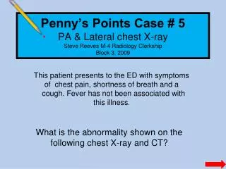
Download
1 / 6
60 likes | 87 Vues
Explore the features of cardiomegaly on chest X-ray, distinguishing various etiologies like cardiomyopathy, congenital heart disease, and valvular disorders. Discover the diagnostic significance of cardio-thoracic ratios and cardiac shadows.

E N D
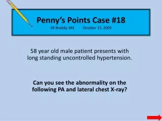
Penny’s Points Case #18Jill Braddy, M4 October 15, 2009 58 year old male patient presents with long standing uncontrolled hypertension. Can you see the abnormality on the following PA and lateral chest X-ray?
? ?
Compare with normal chest normal
The cardio-thoracic ratio is less than 50 percent The cardiac shadow should not extend over over the spine on a lateral chest x-ray.
CardiomegalyHeart enlargement due to dilation or hypertrophy An enlarged cardiac outline (silhouette) can be due to cardiomyopathy, congenital heart disease, valvular heart disease, masses, and pericardial effusions. These etiologies can be somewhat differentiated by chest x-ray. Cardiomyopathy and effusions tend to cause enlargement symmetrically. Cardiac ultrasound / echo is often needed to diagnose pericardial effusion. Congenital heart disease and valvular heart disease will usually effect one chamber. Myopathies can be subdivided into hypertrophic, dilated, and restrictive. Dilated cardiomyopathies result from infection, metabolic disorders, alcohol, and certain chemotherapy drugs. Hypertrophic cardiomyopathy is the most common cause of sudden cardiac death in young athletes. It can be an inherited condition in which the sarcomeres replicate to cause thickening of the myocardium. Restrictive cardiomyopathies are the least common and are usually caused by collagen vascular disorders.