Download
1 / 45
510 likes | 1.68k Vues
Tissues Introduction Epithelial Tissue Classification Glands. Cell Specialization. Multicellularity requires a division of labor Cells look and function differently (specialize) in different parts of the body (ex. bone cell vs. nerve cell)

E N D
Cell Specialization • Multicellularity requires a division of labor • Cells look and function differently (specialize) in different parts of the body (ex. bone cell vs. nerve cell) • Cells specialize into types of tissues, then form organs.
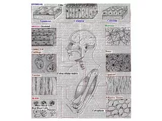
Histology Study of tissues (groups of cells that are similar in structure and function) 4 Tissue Types
1. Connective Tissue • Support • Connect layers of tissue to each other • Bone, ligaments, fat (we will study these in great detail in class later…)
2. Nervous Tissue • Control • Brain, nerves, spinal cord • Highly specialized cells that generate and conduct nerve impulses
3. Muscular Tissue • Movement • Highly vascular • Contracts with cytoskeletal microfilament (actin) that works with motor protein called myosin • 2 categories: • Striated muscle tissue (voluntary or indirect voluntary control) • Smooth muscle tissue (involuntary)
3. Muscular Tissue • Striated Muscle • Ex. Skeletal • Attached to skeletal bones • Long, cylindrical multi-nucleited cells (as in background) • Visible striations • Voluntary control • Ex. Cardiac • Also striated, but uni-nucleated • Branching cells fit tightly with special junctions called intercalated discs
3. Muscular Tissue • Smooth muscle • No striations • Spindle shaped • One central nucleus • Involuntary muscle • Ex. digestive system
4. Epithelial Tissue Interface tissue that forms boundaries between environments and lines surfaces “epithe-” means “laid on” • Coverings and Protection (ex. skin, serous membranes) • Excretion & Secretion (ex. glands) • Filtration (ex. kidneys) • Absorption (ex. digestive system)
1. Tight fitting sheets Regardless of cell shape or number of layers Identifying Characteristics of Epithelial Tisues
2. Apical-Basal Polarity Apical Surface = top surface that borders an “open” space called LUMEN Basal Surface = bottom surface that borders underlying supportive connective tissue Identifying Characteristics LUMEN Connective Tissue
Apical Surface Often w/ microvilli (brush border) Increases SA in areas that need to absorb or secrete Some with cilia (longer) to move substances along lumen Identifying Characteristics LUMEN Connective Tissue
Basal Surface Has adhesive sheet of glyco-proteins secreted by epithelial cells called the basal lamina Connective Tissue beneath secretes collagen, creating the Reticular Lamina. Basal Lamina + Reticular Lamina = Basement Membrane (defines the epithelial boundary) Identifying Characteristics LUMEN LUMEN Connective Tissue
3. Avascular (a = without) Lacks blood vessels Nourished by connective tissue But Innervated w/ nerve fibers 4. Regeneration and repair quickly Identifying Characteristics
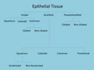
Classification of Epithelial Tissue:Cell Shape Cross-section • Squamous – flat, like a fried egg, or scale-like • Cuboidal – cubes, large spherical central nuclei • Columnar – columns, long oval nuclei, usually near basal surface
Simple (one layer) Thin: limited, no protection Sparse cytoplasm Found where rapid diffusion is a priority (ex. kidneys, lungs) Stratified (many layers) Thick Protective role, subject to wear and tear Regenerate from basal surface to replace apical surface cells that rub off or die Cells differ in shape at apical and basal surface. (named for apical surface) Classification of Epithelial Tissue Cell Layers
Pseudo-stratified false • Shapes vary in height • Nuclei at different levels – appear stratified, but aren’t. • All cells reach basement membrane; only a few reach the surface
Simple Squamous Epithelium One layer Flat Function and Location • Areas of high diffusion rates: • gasses (ex lungs) • nutrients and waste exchange (blood vessels and surrounding cells) • filtrates (kidneys) • Makes lubricating fluid in lining of body cavities (ex. serous membranes)
Simple Squamous Epithelium(Top View) – cells fit like tiled floor
Simple Squamous Epithelium(side view/cross section) – cells look like fried egg LUMEN Figure 4.2
Kidney LUMEN Nucleus of squamous cell
Simple Cuboidal Epithelium Cubed One layer Function and Location • Secretion and Absorption • Covers walls of SMALL ducts, glands, kidney tubules, ovaries
Cuboidal Cell Spherical, large nuclei LUMEN Basement membrane Apical surface
Basal surface Spherical, large nuclei LUMEN Apical surface
Simple Columnar Epithelium One layer columns • Function and Location • Absorption & Secretion (ex. digestive tract) • When in open to body cavities – called mucous membranes • Special Features • Often w/ microvilli on apical surface (brush border) • Goblet cells, single cell glands, produce protective mucus.
Basal surface Apical surface LUMEN
Pseudostratified Epithelium • Function • Absorption • Secretion of mucus by goblet cells • Cilia (larger than microvilli) sweep mucus • Location • Respiratory Linings & Reproductive tract
LUMEN Cilia Basement Membrane Multilevel nuclei
Stratified Squamous Epithelium Multi-layer (thick!) Flat (only cells on apical surface) • Structure • Cells often cuboidal or columnar below apical squamous layer • Function and Location • Protection • Keratin (protein) is accumulated in older cells near the surface – waterproofs and toughens skin • Location • Skin (keratinized), mouth & throat
keratin Squamous Cuboidal Columnar Basement Membrane Dense-Irregular Connective Tissue
Transitional Epithelium • Structure • Multi-layer • Basal surface cells are cuboidal or columnar • Apical surface cells vary: changes shape to accommodate for change in volume due to stretching • Function • Allows stretching • Location • Urinary bladder, ureters & urethra
Glands • Cells that secrete or export a product. • Secretion = protein, lipids, hormones, steroids, acids • Endocrine glands (internally secreting) • No duct, release secretion into blood vessels • Often hormones • Thyroid, adrenal and pituitary glands • Exocrine glands (externally secreting) • Contain ducts, empty onto epithelial surface • Sweat, Oil glands, Salivary glands, Mammary glands.
Shapes of Exocrine glands • Branching of ducts • Simple – single, unbranched duct • Compound – branched duct. • Shape of glands: • Tubular – tube - like • Alveolar – flasks or sacs • Tubuloalveolar – has both tubes and sacs in gland
Merocrine Modes of Secretion Merocrine • Released by exocytosis • Gland is not altered (Ex: Sweat glands and salivary glands) Holocrine • Gland ruptures and releases secretion and dead cells as well. • Sebaceous (ex. oil glands on the face) Holocrine