Download
1 / 62
640 likes | 978 Vues
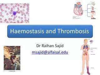
Dr Raihan Sajid msajid@alfaisal.edu. Objectives. Normal haemostasis Functions of vessel wall, platelets and coagulation factors in bleeding, coagulation and thrombosis Clinical correlation in defects of vessel wall, platelets and coagulation factors . Primary haemostasis.

E N D
Dr Raihan Sajid msajid@alfaisal.edu
Objectives • Normal haemostasis • Functions of vessel wall, platelets and coagulation factors in bleeding, coagulation and thrombosis • Clinical correlation in defects of vessel wall, platelets and coagulation factors
Primary haemostasis • Normal physiological response that can prevent blood loss when the vascular system is breached • Inappropriate activation of the system can be very detrimental • System involves coordinated contributions from • Endothelium • Platelets • Plasma proteins
Hemostasis is a balancing act! pro-clotting anti-clotting plugs up holes in blood vessels keeps clotting under control
Pro-Clotting vessels platelets cascade
Pro-Clotting clot
Pro-Clotting • Blood vessel constricts. • Blood loss decreases • Platelets and factors meet
Pro-Clotting • Platelets form a plug. • Proteins are exposed • Platelets adhese • Granules release contents • Platelets aggregate • Phospholipids are exposed
Pro-Clotting • Fibrin seals up plug. • Tissue factor is exposed • Cascade begins • Cascade makes fibrin • Fibrin solidifies plug
Role of endothelium • Intact endothelial cells maintain liquid blood flow by actively inhibiting platelet adherence, preventing coagulation factor activation and lysing blood clot that may form • Endothelial cells can be stimulated by direct injury or by various cytokines that are produced during inflammation.
Role of endothelium • Stimulation results in expression of procoaluant proteins e.g. tissue factor and vWf that contribute to local thrombus formation • Loss of endothelial integrity exposes underlying vWF and basement membrane collagen both substrates for platelet aggregation and thrombus formation
Role of platelets • Two types of granules • α granules • Express the adhesion molecule P-selectin and contain fibrinogen, vWF, factor V and VIII, PF4, PDGF and TGF-α • Dense bodies • Contain ADP, ATP, Calcium, histamine, serotonin and epinephrine
Role of platelets (cont’d) • After injury platelets come in contact with collagen and vWF and undergo three changes • Adhesion and shape change • Secretion or release reaction • aggregation
Physiology of primary haemostasis A. Adhesion B. Aggregation • Fibrin generation
Platelets become ‘sticky’ and adhere to tissue collagen Contact with damaged vascular surface Platelets release ADP, thromboxane A2 Platelet plug Adhere to platelets in plug More platelets become ‘sticky’
Summary of platelet function • Endothelial injury exposes the underlying basement membrane ECM • Platelets adhere to the ECM and become activated by binding to vWF through Gp1B receptors • Platelets provide a phospholipid surface for coagulation • After activation platelets secrete granule products including calcium that activates coagulation proteins, ADP which mediates further platelet aggregation and TXA2 which increases platelet activation and cause vasoconstriction
Summary of platelet function • Released ADP stimulates formation of primary haemostatic plug by activating GpIIb-IIIa receptors that facilitate fibrinogen binding and cross linking • The formation of definitive secondary plug requires activation of thrombin to cleave fibrinogen and form polymerized fibrin via the coagulation cascade
Clotting/coagulation factors Proteins that are involved in the blood coagulation process Most of them are proteases Fibrin polymerisation fluid jelly
INTRINSIC PATHWAY EXTRINSIC PATHWAY Surface contact Tissue injury XII, XI, IX Tissue factor, VII FINAL COMMON PATHWAY V, phospholipid X Xa II (Prothrombin) Thrombin Fibrinogen Fibrin Coagulation cascade INTRINSIC PATHWAY: Activation requires local damage to the endothelial surface of a blood vessel. Starts from INSIDE blood vessels Factor XII = Hageman factor
INTRINSIC PATHWAY EXTRINSIC PATHWAY Surface contact Tissue injury XII, XI, IX Tissue factor, VII FINAL COMMON PATHWAY V, phospholipid X Xa II (Prothrombin) Thrombin Fibrinogen Fibrin Coagulation cascade EXTRINSIC PATHWAY: Activation requires tissue injury. Starts from blood vessel injury and release of tissue factor
INTRINSIC PATHWAY EXTRINSIC PATHWAY Surface contact Tissue injury XII, XI, IX Tissue factor, VII FINAL COMMON PATHWAY Factor V, platelet phospholipid X Xa II (Prothrombin) Thrombin Fibrinogen Fibrin Coagulation cascade
Where does tissue factor come from? • Endothelial cells and monocytes (during inflammation) • It is also present on skin, organ surfaces, epithelial-mesenchymal surfaces.
tissue factor fibrin clot
Role of thrombin in hemostasis and cellular activation. Thrombin plays a critical role in generating cross-linked fibrin (by cleaving fibrinogen to fibrin, and by activating factor XIII), as well as activating several other coagulation factors (see Fig. 4-8). Through protease-activated receptors (PARs, see text), thrombin also modulates several cellular activities. It directly induces platelet aggregation and TxA2 production, and activates ECs to express adhesion molecules, and a variety of fibrinolytic (t-PA), vasoactive (NO, PGI2), and cytokine mediators (e.g., PDGF). Thrombin also directly activates leukocytes. ECM, extracellular matrix; NO, nitric oxide; PDGF, platelet-derived growth factor; PGI2, prostacyclin; TxA2, thromboxane A2; t-PA, tissue plasminogen activator. See Figure 4-7 for additional anticoagulant activities mediated by thrombin, including via thrombomodulin.
PT INR and APTT • PT INR measures the extrinsic and common pathway • Factors II,V,VII,X and fibrinogen • APTT measure intrinsic and common pathway • Fibrinogen, II, V, VIII, IX, X, XI, XII
PT INR • To do a PT (or INR), you just take the patient's plasma (from which the calcium has been removed), and add thromboplastin (as well as calcium). Thromboplastin is just a combination of phospholipid and something that acts like tissue factor. Adding thromboplastin to the patient's plasma will activate the extrinsic pathway. Then you just measure the time (in seconds) it takes to make fibrin.
PT INR • to monitor patients on Coumadin (warfarin). • to evaluate a patient who is in liver failure (in whom all the coagulation factors would decline) • patient in disseminated intravascular coagulation (who is using up all his or her coag factors) • Factor VII deficiency • as part of a pre-op screening panel.
APTT • To do APTT, you take the patient's plasma (from which the calcium has been removed), and add phospholipid (as well as calcium). That's actually why this test is called the "partial" thromboplastin time...when they first did this assay, they didn't know what was in thromboplastin. But they did know that if you just added part of the thromboplastin substance, you could get the intrinsic pathway to run (hence the name "partial" thromboplastin time).Someone figured out later that thromboplastin was a combination of phospholipid and something that acts like tissue factor...and the part of thromboplastin that was being added in the PTT was just the phospholipid part.
APTT • to monitor patients on heparin • factor VIII and IX deficiency • to evaluate a patient who is in liver failure (in whom all the coagulation factors would decline) • patient in disseminated intravascular coagulation (who is using up all his or her coagulation factors) • as part of a pre-op screening panel.
Anti-Clotting clot
Anti-Clotting clot • 1 • cascade inhibition • TFPI • ATIII • Proteins C, S
Anti-Clotting clot • 1 • cascade inhibition • TFPI • ATIII • Proteins C, S • 2 • clot lysis • t-PA • plasmin
Fibrinolysis (Plasminogen activator inhibitor 1 And alpha 2 antiplasmin inhibits t-PA) - Tissue from endothelium etc plasminogen activator (t-PA) digests Fibrin & other coagulants plasmin plasminogen FDPs = Fibrin degradation products
Intrinsic Extrinsic TFPI ATIII protein C thrombin exposed TF XI XIa TF VIIa IX IXa VII VIIIa VIII X Xa Va V prothrombin thrombin clot fibrinogen fibrin
Clinical correlation • Problems with platelets • Problems with coagulation cascade • 10 minute break
Thrombosis Haemostasis is normally beneficial BUT if inappropriately activated can be harmful Three predisposing situations/factors - Known as Virchow’s triad • Changes in the intimal surface of the vessel • Changes in the pattern of blood flow • Changes in the blood constituents
Distinguish between these words • Thrombosis (pathological) • A solid mass of blood constituents [thrombus] formed within the a vascular lumen during life • Clot • A solid mass of blood constituents occurring outside the vascular tree OR after death within the vascular tree
Endothelial injury • Endothelial injury is particularly important for thrombus formation in the heart or the arterial circulation, where the normally high flow rates might otherwise impede clotting by preventing platelet adhesion and washing out activated coagulation factors. • Thus, thrombus formation within cardiac chambers (e.g., after endocardial injury due to myocardial infarction), over ulcerated plaques in atherosclerotic arteries, or at sites of traumatic or inflammatory vascular injury (vasculitis) is largely a consequence of endothelial cell injury. • Any disturbance in the dynamic balance of the prothombotic and antithrombotic activities of endothelium can influence local clotting events
Endothelial injury (cont’d) • Thus, dysfunctional endothelial cells can produce more procoagulant factors (e.g., platelet adhesion molecules, tissue factor, PAIs) or may synthesize less anticoagulant effectors (e.g., thrombomodulin, PGI2, t-PA). • Endothelial dysfunction can be induced by a wide variety of insults, including • hypertension • turbulent blood flow • bacterial endotoxins • radiation injury • homocystinemia or hypercholesterolemia, • toxins absorbed from cigarette smoke.
Alteration in blood flow • Turbulence contributes to arterial and cardiac thrombosis by causing endothelial injury or dysfunction, as well as by forming countercurrents and local pockets of stasis • Stasis is a major contributor in the development of venous thrombi • Normal blood flow is laminarsuch that the platelets (and other blood cellular elements) flow centrally in the vessel lumen, separated from endothelium by a slower moving layer of plasma
Stasis and turbulence leads to • endothelial activation, enhancing pro-coagulant activity and leukocyte adhesion • Disrupt laminar flow and bring platelets into contact with the endothelium • Prevent washout and dilution of activated clotting factors by fresh flowing blood and the inflow of clotting factor inhibitors